Koser David E, Moeendarbary Emad, Hanne Janina, Kuerten Stefanie, Franze Kristian
Department of Physiology, Development and Neuroscience, University of Cambridge, Cambridge, United Kingdom; Department of Anatomy I, University of Cologne, Cologne, Germany.
Department of Physiology, Development and Neuroscience, University of Cambridge, Cambridge, United Kingdom.
Biophys J. 2015 May 5;108(9):2137-47. doi: 10.1016/j.bpj.2015.03.039.
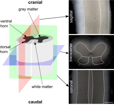
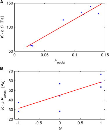
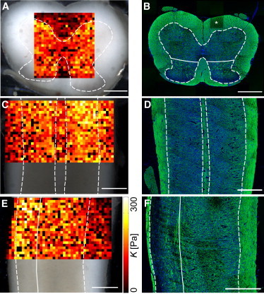
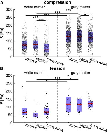
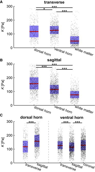
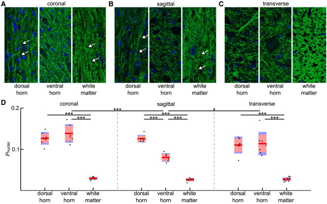
Mechanical signaling plays an important role in cell physiology and pathology. Many cell types, including neurons and glial cells, respond to the mechanical properties of their environment. Yet, for spinal cord tissue, data on tissue stiffness are sparse. To investigate the regional and direction-dependent mechanical properties of spinal cord tissue at a spatial resolution relevant to individual cells, we conducted atomic force microscopy (AFM) indentation and tensile measurements on acutely isolated mouse spinal cord tissue sectioned along the three major anatomical planes, and correlated local mechanical properties with the underlying cellular structures. Stiffness maps revealed that gray matter is significantly stiffer than white matter irrespective of directionality (transverse, coronal, and sagittal planes) and force direction (compression or tension) (K(g) = ∼ 130 P(a) vs. K(w) = ∼ 70 Pa); both matters stiffened with increasing strain. When all data were pooled for each plane, gray matter behaved like an isotropic material under compression; however, subregions of the gray matter were rather heterogeneous and anisotropic. For example, in sagittal sections the dorsal horn was significantly stiffer than the ventral horn. In contrast, white matter behaved transversely isotropic, with the elastic stiffness along the craniocaudal (i.e., longitudinal) axis being lower than perpendicular to it. The stiffness distributions we found under compression strongly correlated with the orientation of axons, the areas of cell nuclei, and cellular in plane proximity. Based on these morphological parameters, we developed a phenomenological model to estimate local mechanical properties of central nervous system (CNS) tissue. Our study may thus ultimately help predicting local tissue stiffness, and hence cell behavior in response to mechanical signaling under physiological and pathological conditions, purely based on histological data.
机械信号在细胞生理和病理过程中发挥着重要作用。包括神经元和神经胶质细胞在内的许多细胞类型都会对其周围环境的力学特性做出反应。然而,对于脊髓组织,关于组织硬度的数据却很稀少。为了在与单个细胞相关的空间分辨率下研究脊髓组织的区域和方向依赖性力学特性,我们对沿三个主要解剖平面切片的急性分离小鼠脊髓组织进行了原子力显微镜(AFM)压痕和拉伸测量,并将局部力学特性与潜在的细胞结构相关联。硬度图显示,无论方向(横向、冠状面和矢状面)和力的方向(压缩或拉伸)如何,灰质都明显比白质硬(K(g) = ∼ 130 P(a) 对 K(w) = ∼ 70 Pa);两种组织都随着应变增加而变硬。当将每个平面的所有数据汇总时,灰质在压缩下表现得像各向同性材料;然而,灰质的子区域相当不均匀且各向异性。例如,在矢状切片中,背角比腹角明显更硬。相比之下,白质表现为横向各向同性,沿着头尾(即纵向)轴的弹性刚度低于与其垂直的方向。我们在压缩下发现的硬度分布与轴突的方向、细胞核的面积以及细胞在平面内的接近程度密切相关。基于这些形态学参数,我们开发了一个唯象模型来估计中枢神经系统(CNS)组织的局部力学特性。因此,我们的研究最终可能有助于仅基于组织学数据预测局部组织硬度,从而预测生理和病理条件下细胞对机械信号的反应行为。