Lacoste Alix M B, Schoppik David, Robson Drew N, Haesemeyer Martin, Portugues Ruben, Li Jennifer M, Randlett Owen, Wee Caroline L, Engert Florian, Schier Alexander F
Department of Molecular and Cellular Biology, Harvard University, Cambridge, MA 02138, USA.
Program in Neuroscience, Harvard Medical School, Boston, MA 02115, USA.
Curr Biol. 2015 Jun 1;25(11):1526-34. doi: 10.1016/j.cub.2015.04.025. Epub 2015 May 7.
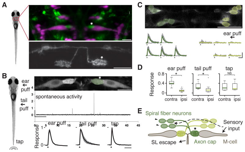
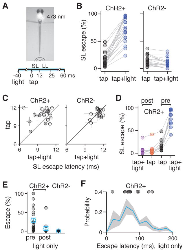
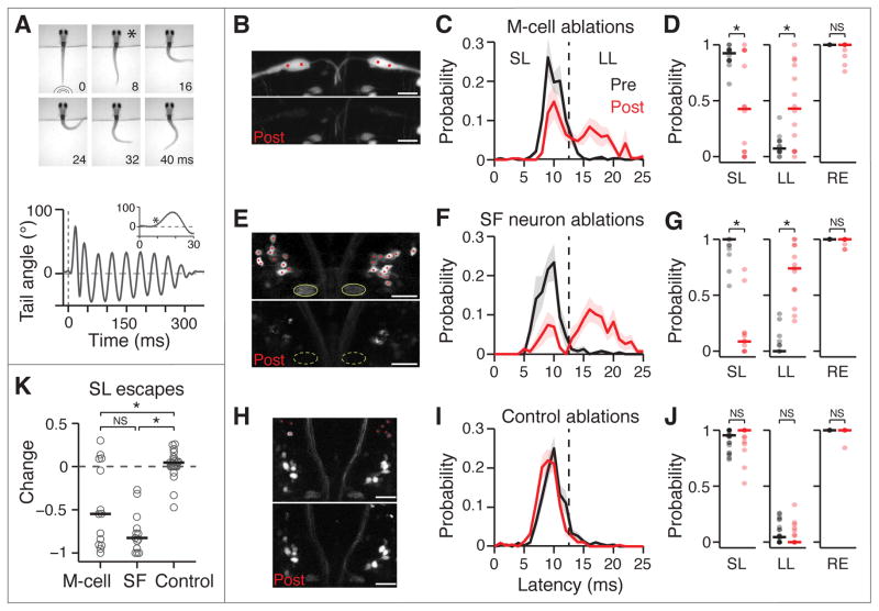
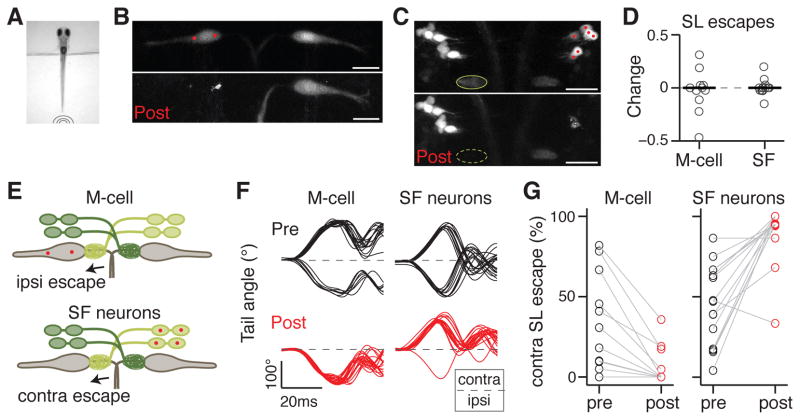
The Mauthner cell (M-cell) is a command-like neuron in teleost fish whose firing in response to aversive stimuli is correlated with short-latency escapes [1-3]. M-cells have been proposed as evolutionary ancestors of startle response neurons of the mammalian reticular formation [4], and studies of this circuit have uncovered important principles in neurobiology that generalize to more complex vertebrate models [3]. The main excitatory input was thought to originate from multisensory afferents synapsing directly onto the M-cell dendrites [3]. Here, we describe an additional, convergent pathway that is essential for the M-cell-mediated startle behavior in larval zebrafish. It is composed of excitatory interneurons called spiral fiber neurons, which project to the M-cell axon hillock. By in vivo calcium imaging, we found that spiral fiber neurons are active in response to aversive stimuli capable of eliciting escapes. Like M-cell ablations, bilateral ablations of spiral fiber neurons largely eliminate short-latency escapes. Unilateral spiral fiber neuron ablations shift the directionality of escapes and indicate that spiral fiber neurons excite the M-cell in a lateralized manner. Their optogenetic activation increases the probability of short-latency escapes, supporting the notion that spiral fiber neurons help activate M-cell-mediated startle behavior. These results reveal that spiral fiber neurons are essential for the function of the M-cell in response to sensory cues and suggest that convergent excitatory inputs that differ in their input location and timing ensure reliable activation of the M-cell, a feedforward excitatory motif that may extend to other neural circuits.
莫氏细胞(M细胞)是硬骨鱼体内一种类似指令的神经元,其对厌恶刺激的放电与短潜伏期逃避反应相关[1-3]。M细胞被认为是哺乳动物网状结构惊吓反应神经元的进化祖先[4],对该神经回路的研究揭示了神经生物学中的重要原理,这些原理可推广到更复杂的脊椎动物模型[3]。人们认为主要的兴奋性输入源自多感觉传入神经,它们直接与M细胞的树突形成突触[3]。在此,我们描述了一条额外的、汇聚性通路,它对于斑马鱼幼体中M细胞介导的惊吓行为至关重要。该通路由称为螺旋纤维神经元的兴奋性中间神经元组成,它们投射到M细胞的轴丘。通过活体钙成像,我们发现螺旋纤维神经元在对能够引发逃避的厌恶刺激做出反应时会被激活。与M细胞消融一样,双侧消融螺旋纤维神经元在很大程度上消除了短潜伏期逃避反应。单侧消融螺旋纤维神经元会改变逃避的方向性,这表明螺旋纤维神经元以一种偏向化的方式兴奋M细胞。它们的光遗传学激活增加了短潜伏期逃避反应的概率,支持了螺旋纤维神经元有助于激活M细胞介导的惊吓行为这一观点。这些结果表明,螺旋纤维神经元对于M细胞响应感觉线索的功能至关重要,并表明在输入位置和时间上不同的汇聚性兴奋性输入可确保M细胞的可靠激活,这是一种前馈兴奋性基序,可能延伸到其他神经回路。