Crisostomo Veronica, Baez-Diaz Claudia, Maestre Juan, Garcia-Lindo Monica, Sun Fei, Casado Javier G, Blazquez Rebeca, Abad Jose L, Palacios Itziar, Rodriguez-Borlado Luis, Sanchez-Margallo Francisco M
Jesús Usón Minimally Invasive Surgery Centre, Carretera N-521, km 41.8, 10071, Cáceres, Spain.
Coretherapix, Santiago Grisolía, n° 2 Parque Científico de Madrid, 28760, Tres Cantos, Madrid, Spain.
J Transl Med. 2015 May 12;13:156. doi: 10.1186/s12967-015-0512-2.
The optimal timing of cardiac stem cells administration is still unclear. We assessed the safety of same-day and delayed (one week) delivery and the possible influence of the timing on the therapeutic outcomes of allogeneic porcine cardiac stem cells administration after acute myocardial infarction in a closed-chest ischemia-reperfusion model.
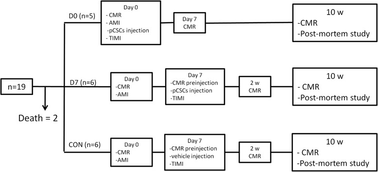
Female swine surviving 90 min occlusion of the mid left anterior descending coronary artery received an intracoronary injection of 25x10(6) porcine cardiac stem cells either two hours (n = 5, D0) or 7 days (n = 6, D7) after reperfusion. Controls received intracoronary injection of vehicle on day 7 (n = 6, CON). Safety was defined in terms of absence of major cardiac events, changes to the ECG during injection, post-administration coronary flow assessed using the TIMI scale and cardiac troponin I determination after the intervention. Cardiac Magnetic Resonance was performed for morphological and functional assessment prior to infarction, before injection (D7 and CON groups only), at one and 10 weeks. Samples were taken from the infarct and transition areas for pathological examination.
No major adverse cardiac events were seen during injection in any group. Animals receiving the therapy on the same day of infarction (D0 group) showed mild transient ST changes during injection (n = 4) and, in one case, slightly compromised coronary flow (TIMI 2). Cardiac function parameters and infarct sizes were not significantly different between groups, with a trend towards higher ejection fraction in the treated groups. Ventricular volumes indexed to body surface area increased over time in control animals, and decreased by the end of the study in animals receiving the therapy, significantly so when comparing End Diastolic Volume between CON and D7 groups (CON: 121.70 ml/m(2) ± 26.09 ml/m(2), D7: 98.71 ml/m(2) ± 8.30 ml/m(2), p = 0.037). The treated groups showed less organization of the collagenous scar, and a significantly (p = 0.019) higher amount of larger, more mature vessels at the infarct border.
The intracoronary injection of 25x10(6) allogeneic cardiac stem cells is generally safe, both early and 7 days after experimental infarction, and alleviates myocardial dysfunction, with a greater limitation of left ventricular remodeling when performed at one week.
心脏干细胞给药的最佳时机仍不明确。我们在闭胸缺血再灌注模型中评估了急性心肌梗死后当日和延迟(一周)给药的安全性,以及给药时机对同种异体猪心脏干细胞治疗效果的可能影响。
在左前降支冠状动脉中段闭塞90分钟后存活的雌性猪,在再灌注后2小时(n = 5,D0组)或7天(n = 6,D7组)接受冠状动脉内注射25×10⁶个猪心脏干细胞。对照组在第7天接受冠状动脉内注射赋形剂(n = 6,CON组)。安全性通过无重大心脏事件、注射期间心电图变化、使用TIMI量表评估给药后冠状动脉血流以及干预后测定心肌肌钙蛋白I来定义。在梗死前、注射前(仅D7组和CON组)、1周和10周时进行心脏磁共振成像以进行形态学和功能评估。从梗死和过渡区域采集样本进行病理检查。
任何组在注射期间均未观察到重大不良心脏事件。在梗死当日接受治疗的动物(D0组)在注射期间显示轻度短暂ST段改变(n = 4),且有1例冠状动脉血流略有受损(TIMI 2级)。各组之间的心脏功能参数和梗死面积无显著差异,治疗组的射血分数有升高趋势。以体表面积指数衡量的心室容积在对照动物中随时间增加,而在接受治疗的动物中在研究结束时减小,比较CON组和D7组的舒张末期容积时差异显著(CON组:121.70 ml/m²±26.09 ml/m²,D7组:98.71 ml/m²±8.30 ml/m²,p = 0.037)。治疗组的胶原瘢痕组织较少,梗死边界处较大、更成熟血管的数量显著更多(p = 0.019)。
冠状动脉内注射25×10⁶个同种异体心脏干细胞在实验性梗死后早期和7天均普遍安全,可减轻心肌功能障碍,在一周时进行时对左心室重构的限制更大。