Rao Thapi D, Tian Huasong, Ma Xun, Yan Xiujun, Thapi Sahityasri, Schultz Nikolaus, Rosales Nestor, Monette Sebastien, Wang Amy, Hyman David M, Levine Douglas A, Solit David, Spriggs David R
Department of Medicine, Memorial Sloan Kettering Cancer Center; and Department of Medicine, Weill Cornell Medical College, New York, NY, United States of America.
Computational Biology Center, Memorial Sloan Kettering Cancer Center, New York, NY, United States of America.
PLoS One. 2015 May 12;10(5):e0126633. doi: 10.1371/journal.pone.0126633. eCollection 2015.
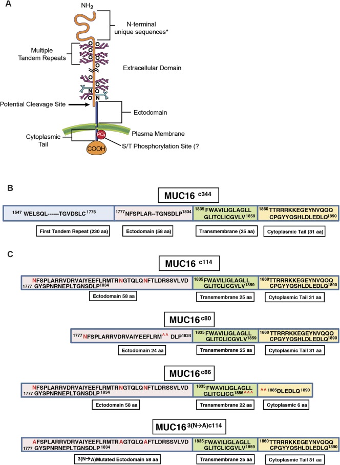
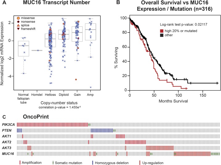
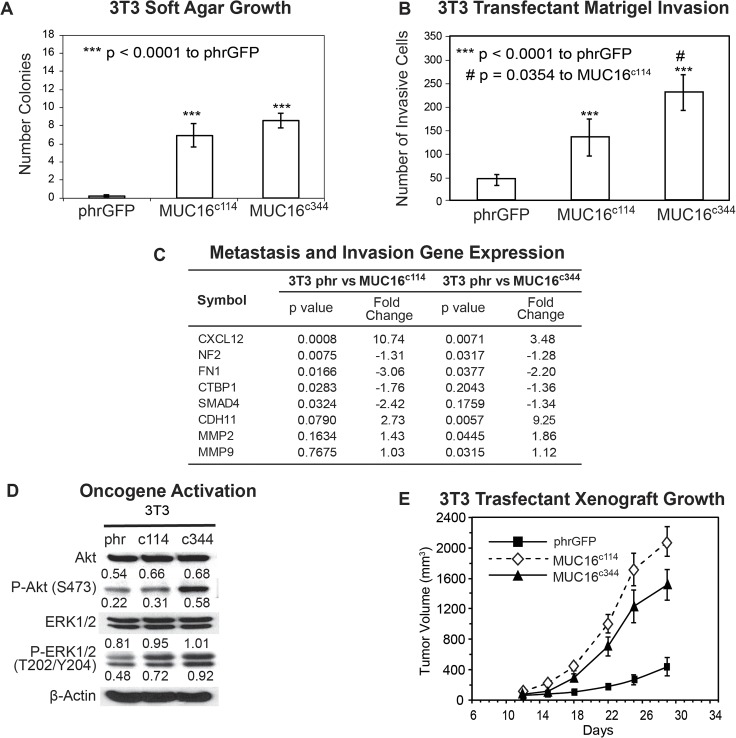
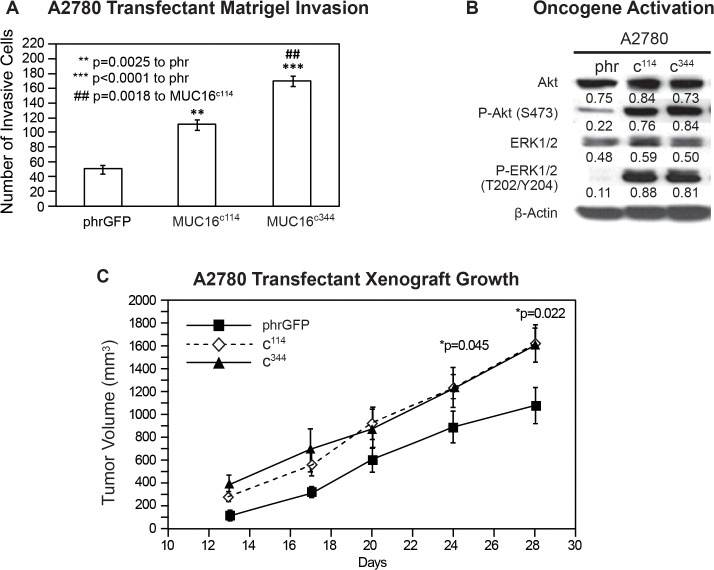
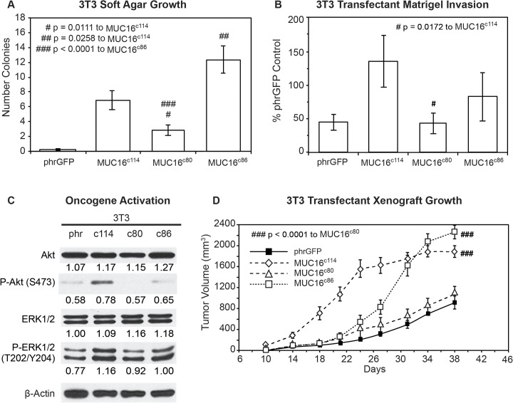
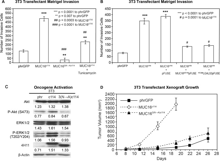
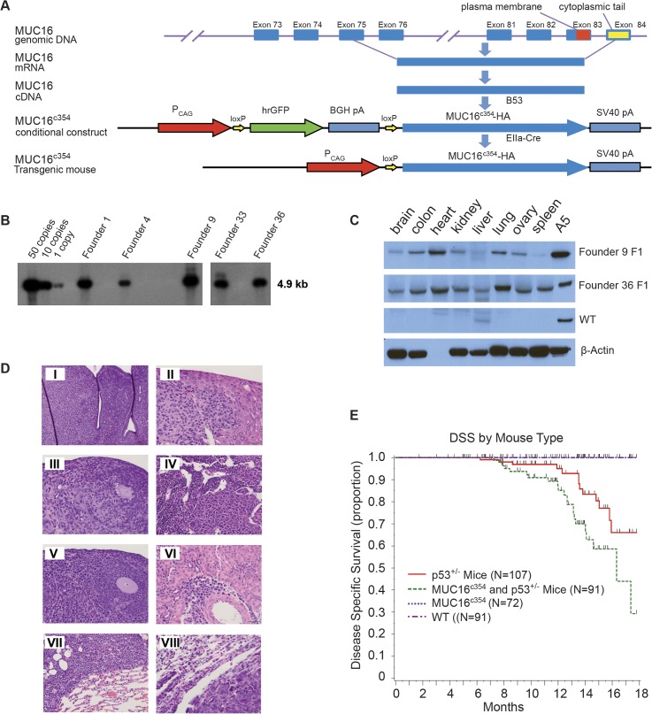
The CA125 antigen is found in the serum of many patients with serous ovarian cancer and has been widely used as a disease marker. CA125 has been shown to be an independent factor for clinical outcome in this disease. In The Cancer Genome Atlas ovarian cancer project, MUC16 expression levels are frequently increased, and the highest levels of MUC16 expression are linked to a significantly worse survival. To examine the biologic effect of the proximal portion of MUC16/CA125, NIH/3T3 (3T3) fibroblast cell lines were stably transfected with the carboxy elements of MUC16. As few as 114 amino acids from the carboxy-terminal portion of MUC16 were sufficient to increase soft agar growth, promote matrigel invasion, and increase the rate of tumor growth in athymic nude mice. Transformation with carboxy elements of MUC16 was associated with activation of the AKT and ERK pathways. MUC16 transformation was associated with up-regulation of a number of metastases and invasion gene transcripts, including IL-1β, MMP2, and MMP9. All observed oncogenic changes were exclusively dependent on the extracellular "ectodomain" of MUC16. The biologic impact of MUC16 was also explored through the creation of a transgenic mouse model expressing 354 amino acids of the carboxy-terminal portion of MUC16 (MUC16c354). Under a CMV, early enhancer plus chicken β actin promoter (CAG) MUC16c354 was well expressed in many organs, including the brain, colon, heart, kidney, liver, lung, ovary, and spleen. MUC16c354 transgenic animals appear to be viable, fertile, and have a normal lifespan. However, when crossed with p53-deficient mice, the MUC16c354:p53+/- progeny displayed a higher frequency of spontaneous tumor development compared to p53+/- mice alone. We conclude that the carboxy-terminal portion of the MUC16/CA125 protein is oncogenic in NIH/3T3 cells, increases invasive tumor properties, activates the AKT and ERK pathways, and contributes to the biologic properties of ovarian cancer.
CA125抗原在许多浆液性卵巢癌患者的血清中被发现,并已被广泛用作疾病标志物。CA125已被证明是该疾病临床结局的一个独立因素。在癌症基因组图谱卵巢癌项目中,MUC16表达水平经常升高,且MUC16表达的最高水平与显著更差的生存率相关。为了研究MUC16/CA125近端部分的生物学效应,用MUC16的羧基元件稳定转染NIH/3T3(3T3)成纤维细胞系。来自MUC16羧基末端部分的仅114个氨基酸就足以增加软琼脂生长、促进基质胶侵袭,并增加无胸腺裸鼠的肿瘤生长速率。用MUC16的羧基元件进行转化与AKT和ERK途径的激活相关。MUC16转化与包括IL-1β、MMP2和MMP9在内的许多转移和侵袭基因转录本的上调相关。所有观察到的致癌变化完全依赖于MUC16的细胞外“胞外域”。还通过创建表达MUC16羧基末端部分354个氨基酸(MUC16c354)的转基因小鼠模型来探索MUC16的生物学影响。在巨细胞病毒早期增强子加鸡β肌动蛋白启动子(CAG)的控制下,MUC16c354在包括脑、结肠、心脏、肾脏、肝脏、肺、卵巢和脾脏在内的许多器官中均有良好表达。MUC16c354转基因动物似乎是有活力的、可育的,并且具有正常的寿命。然而,当与p53缺陷小鼠杂交时,与单独的p53+/-小鼠相比,MUC16c354:p53+/-后代显示出自发性肿瘤发生的频率更高。我们得出结论,MUC16/CA125蛋白的羧基末端部分在NIH/3T3细胞中具有致癌性,增加肿瘤侵袭特性,激活AKT和ERK途径,并有助于卵巢癌的生物学特性。