Department of Biochemistry and Molecular Biology, University of Nebraska Medical Center, Omaha, NE, United States of America.
Sanguine Diagnostics and Therapeutics Inc. Omaha, NE, United States of America.
PLoS One. 2018 Apr 30;13(4):e0193907. doi: 10.1371/journal.pone.0193907. eCollection 2018.
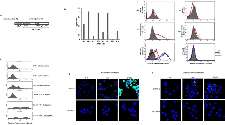
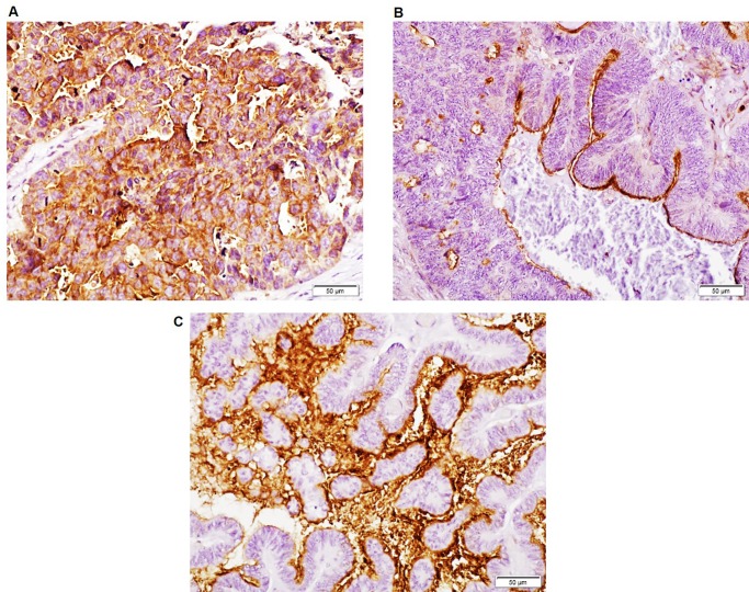
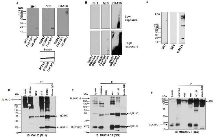
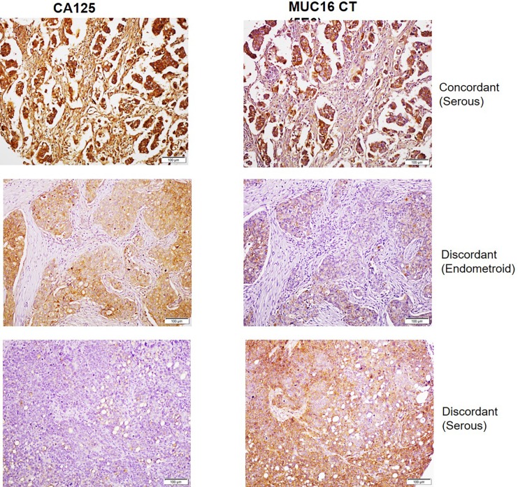
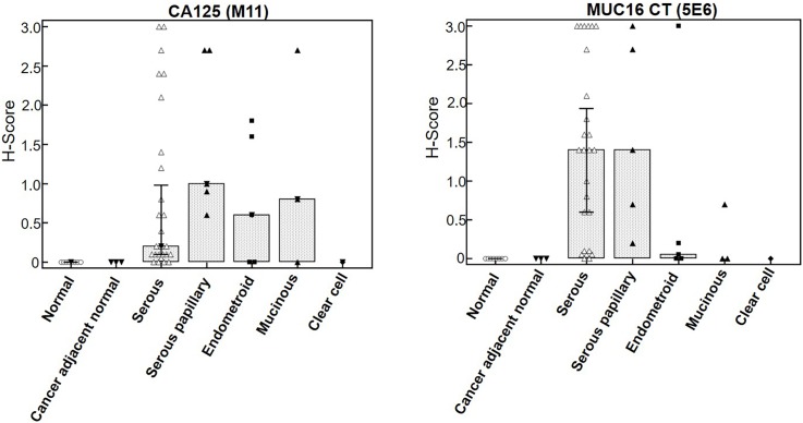
MUC16 is overexpressed in ovarian cancer and plays important roles in invasion and metastasis. Previously described monoclonal antibodies against cell surface expressed MUC16 recognize the N-terminal tandemly repeated epitopes present in cancer antigen 125 (CA125). MUC16 is cleaved at a specific location, thus, releasing CA125 into the extracellular space. Recent reports have indicated that the retained carboxy-terminal (CT) fragment of MUC16 might play an important role in tumorigenicity in diverse types of cancers. However, limited data is available on the fate and existence of CT fragment on the surface of the cancer cell. Herein, we characterize two monoclonal antibodies (mAbs) showing specificity to the retained juxtamembrane region of MUC16. For the first time, we demonstrate that MUC16 is cleaved in ovarian cancer cells (NIH:OVCAR-3 [OVCAR-3]) and that the cleaved MUC16 subunits remain associated with each other. Immunohistochemical analyses on different grades of ovarian tumor tissues indicated differential reactivity of CA125 and MUC16 CT mAbs. The CA125 (M11) mAb detected 32/40 (80%), while the CT mAb (5E6) detected 33/40 (82.5%) of total ovarian cancer cases. For serous and serous papillary cases, the CA125 (M11) mAb stained 27/31 cases (87%), while CT mAb (5E6) stained 29/31 cases (93.5%). The CT mAb(s) accurately predict expression of MUC16 since their epitopes are not tandemly repeated and their reactivity may not be dependent on O-linked glycosylation. These antibodies can serve as valuable reagents for understanding MUC16 cleavage and may also serve as potential therapeutic agents for treatment of ovarian cancer.
MUC16 在卵巢癌中过表达,在侵袭和转移中发挥重要作用。先前描述的针对细胞表面表达的 MUC16 的单克隆抗体识别存在于癌症抗原 125(CA125)中的 N 端串联重复表位。MUC16 在特定位置被切割,从而将 CA125 释放到细胞外空间。最近的报告表明,MUC16 的保留羧基末端(CT)片段可能在多种类型癌症的肿瘤发生中发挥重要作用。然而,关于 CT 片段在癌细胞表面的命运和存在,可用的数据有限。在此,我们鉴定了两种特异性识别 MUC16 保留的跨膜区的单克隆抗体(mAbs)。我们首次证明,MUC16 在卵巢癌细胞(NIH:OVCAR-3 [OVCAR-3])中被切割,并且切割的 MUC16 亚基彼此保持关联。对不同分级的卵巢肿瘤组织的免疫组织化学分析表明,CA125 和 MUC16 CT mAb 的反应性不同。CA125(M11)mAb 检测到 40 例中的 32 例(80%),而 CT mAb(5E6)检测到 40 例中的 33 例(82.5%)。对于浆液性和浆液乳头状病例,CA125(M11)mAb 染色 31 例中的 27 例(87%),而 CT mAb(5E6)染色 31 例中的 29 例(93.5%)。CT mAb 能够准确预测 MUC16 的表达,因为它们的表位不是串联重复的,并且它们的反应性可能不依赖于 O 连接的糖基化。这些抗体可以作为理解 MUC16 切割的有价值的试剂,也可以作为治疗卵巢癌的潜在治疗剂。