Han Chang Yeob, Lim Sang Woo, Koo Ja Hyun, Kim Won, Kim Sang Geon
College of Pharmacy and Research Institute of Pharmaceutical Sciences, Seoul National University, Seoul, Korea.
Department of Internal Medicine, Seoul Metropolitan Government Seoul National University Boramae Medical Center, Seoul, Korea.
Gut. 2016 Aug;65(8):1377-88. doi: 10.1136/gutjnl-2014-308506. Epub 2015 May 12.
Endoplasmic reticulum (ER) stress is involved in liver injury, but molecular determinants are largely unknown. This study investigated the role of pleckstrin homology-like domain, family A, member-3 (PHLDA3), in hepatocyte death caused by ER stress and the regulatory basis.
Hepatic PHLDA3 expression was assessed in HCV patients with hepatitis and in several animal models with ER stress. Immunoblottings, PCR, reporter gene, chromatin immunoprecipitation (ChIP) and mutation analyses were done to explore gene regulation. The functional effect of PHLDA3 on liver injury was validated using lentiviral delivery of shRNA.
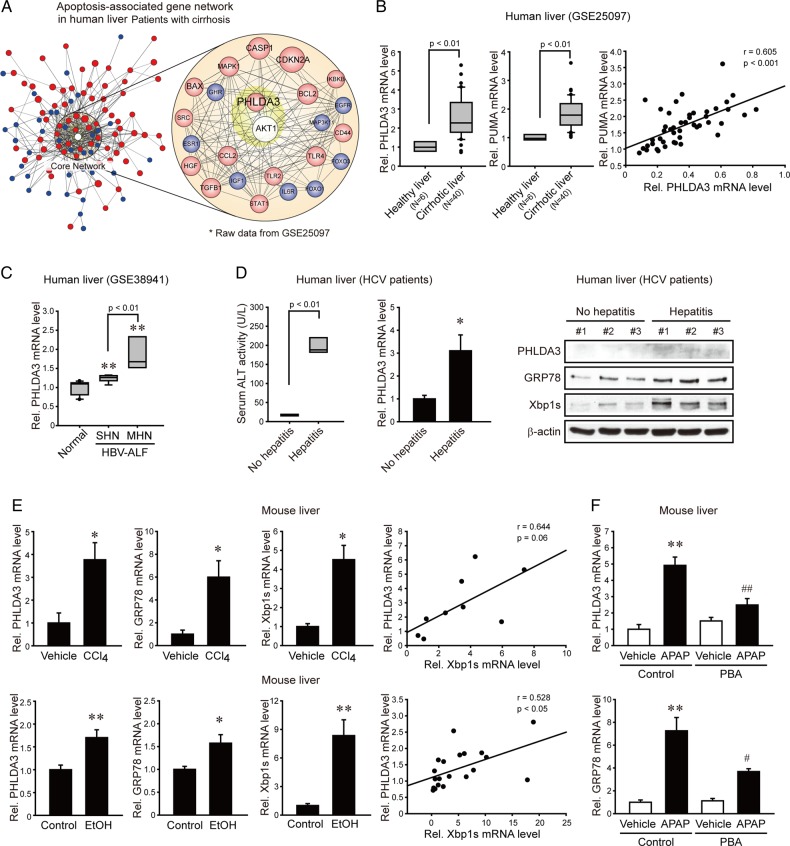
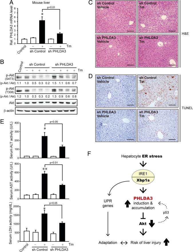
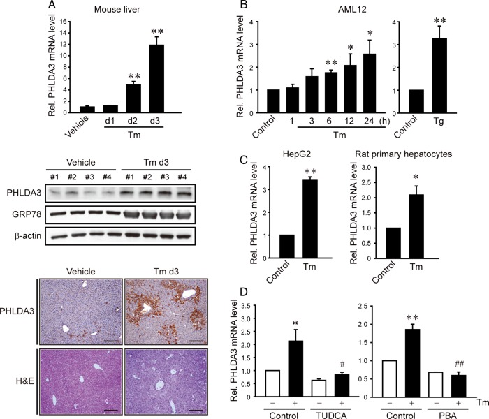
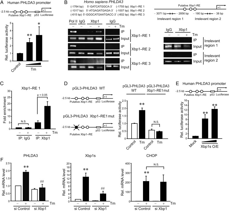
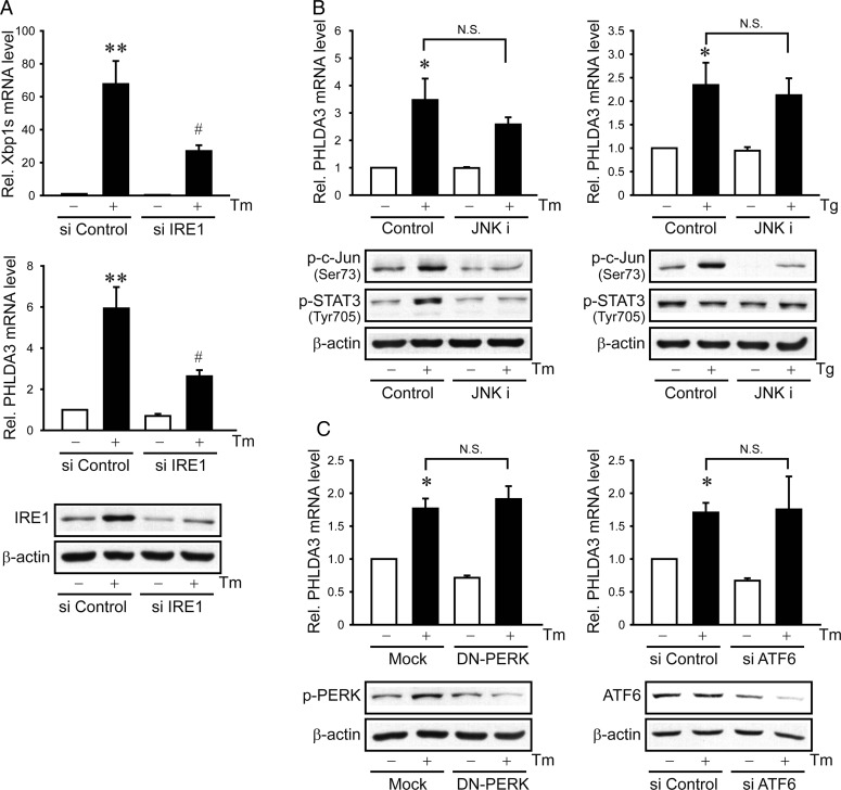
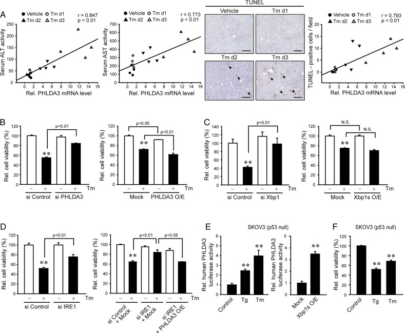
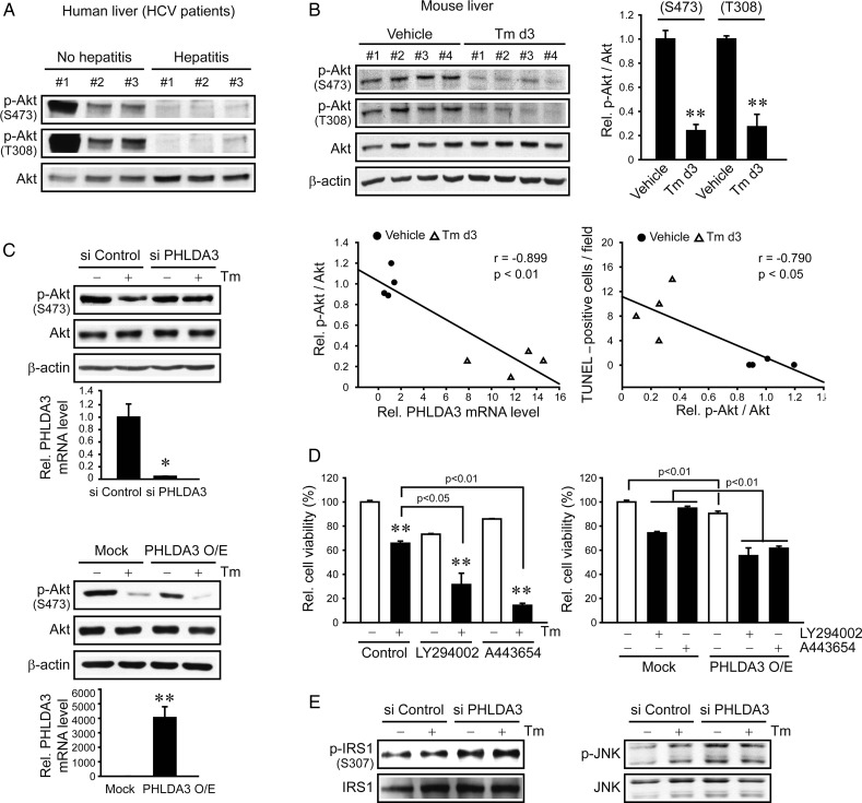
PHLDA3 was overexpressed in relation to hepatocyte injury in patients with acute liver failure or liver cirrhosis or in toxicant-treated mice. In HCV patients with liver injury, PHLDA3 was upregulated in parallel with the induction of ER stress marker. Treatment of mice with tunicamycin (Tm) (an ER stress inducer) increased PHLDA3 expression in the liver. X box-binding protein-1 (Xbp1) was newly identified as a transcription factor responsible for PHLDA3 expression. Inositol-requiring enzyme 1 (IRE1) (an upstream regulator of Xbp1) was required for PHLDA3 induction by Tm, whereas other pathways (c-Jun N-terminal kinase (JNK), protein kinase RNA-like endoplasmic reticulum kinase (PERK) and activating transcription factor 6 (ATF6)) were not. PHLDA3 overexpression correlated with the severity of hepatocyte injury in animal or cell model of ER stress. In p53-deficient cells, ER stress inducers transactivated PHLDA3 with a decrease in cell viability. ER stress-induced hepatocyte death depended on serine/threonine protein kinase B (Akt) inhibition by PHLDA3. Lentiviral delivery of PHLDA3 shRNA to mice abrogated p-Akt inhibition in the liver by Tm, attenuating hepatocyte injury.
ER stress in hepatocytes induces PHLDA3 via IRE1-Xbp1s pathway, which facilitates liver injury by inhibiting Akt.
内质网(ER)应激与肝损伤有关,但分子决定因素大多未知。本研究调查了普列克底物蛋白同源结构域样家族A成员3(PHLDA3)在ER应激引起的肝细胞死亡中的作用及其调控基础。
评估丙型肝炎病毒(HCV)肝炎患者以及几种ER应激动物模型中肝脏PHLDA3的表达。进行免疫印迹、聚合酶链反应(PCR)、报告基因、染色质免疫沉淀(ChIP)和突变分析以探索基因调控。使用慢病毒介导的短发夹RNA(shRNA)验证PHLDA3对肝损伤的功能作用。
在急性肝衰竭或肝硬化患者或经毒物处理的小鼠中,PHLDA3与肝细胞损伤相关而过度表达。在有肝损伤的HCV患者中,PHLDA3与ER应激标志物的诱导同时上调。用衣霉素(Tm,一种ER应激诱导剂)处理小鼠可增加肝脏中PHLDA3的表达。X盒结合蛋白1(Xbp1)被新鉴定为负责PHLDA3表达的转录因子。肌醇需求酶1(IRE1,Xbp1的上游调节因子)是Tm诱导PHLDA3所必需的,而其他途径(c-Jun氨基末端激酶(JNK)、蛋白激酶RNA样内质网激酶(PERK)和活化转录因子6(ATF6))则不是。在ER应激的动物或细胞模型中,PHLDA3的过度表达与肝细胞损伤的严重程度相关。在p53缺陷细胞中,ER应激诱导剂可激活PHLDA3并降低细胞活力。ER应激诱导的肝细胞死亡依赖于PHLDA3对丝氨酸/苏氨酸蛋白激酶B(Akt)的抑制作用。将PHLDA3 shRNA慢病毒导入小鼠可消除Tm对肝脏中磷酸化Akt(p-Akt)的抑制作用,减轻肝细胞损伤。
肝细胞中的ER应激通过IRE1-Xbp1s途径诱导PHLDA3,后者通过抑制Akt促进肝损伤。