Faria Silvana C, Sagebiel Tara, Balachandran Aparna, Devine Catherine, Lal Chandana, Bhosale Priya R
Department of Diagnostic Radiology, MD Anderson Cancer Center, The University of Texas, Houston, Texas, USA.
Department of Diagnostic Radiology, UC Irvine Health, Irvine, California, USA.
Indian J Radiol Imaging. 2015 Apr-Jun;25(2):137-47. doi: 10.4103/0971-3026.155857.
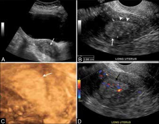
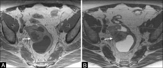
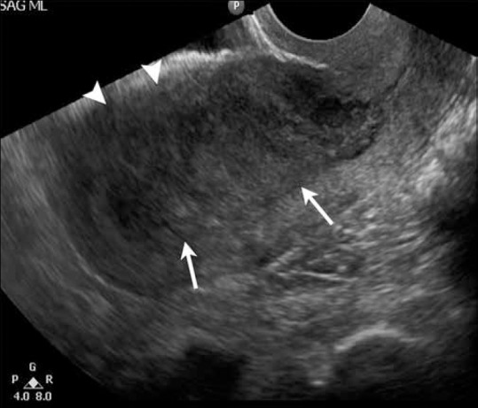
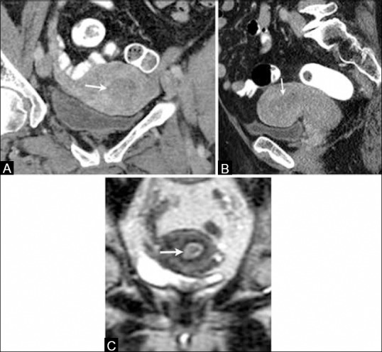
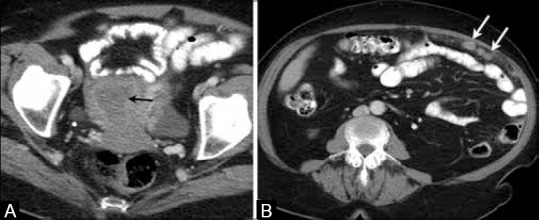
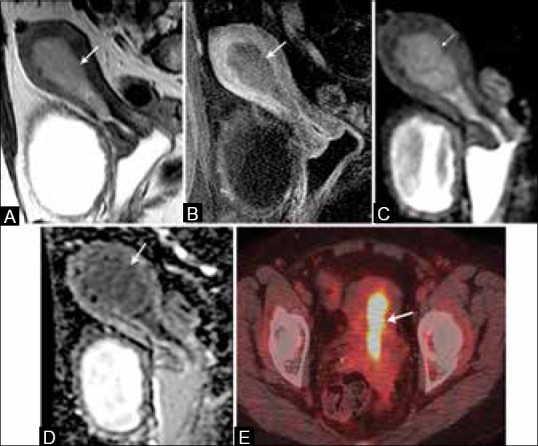
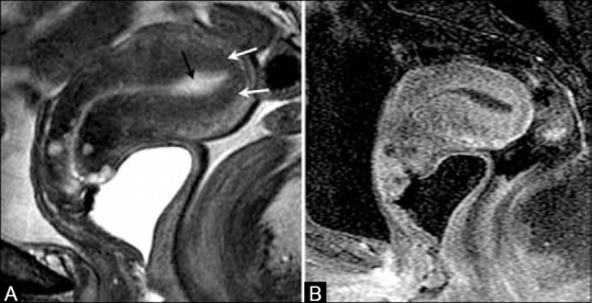
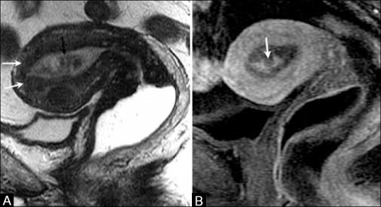
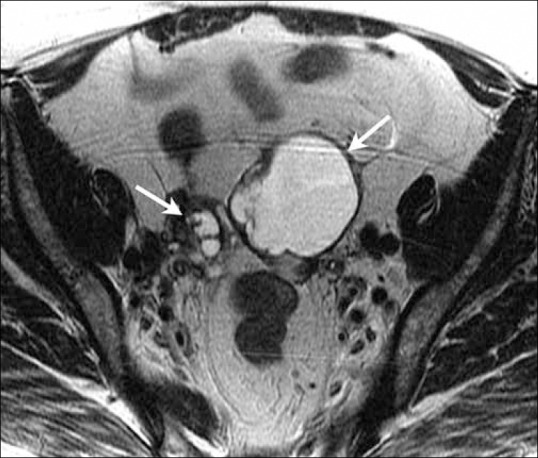
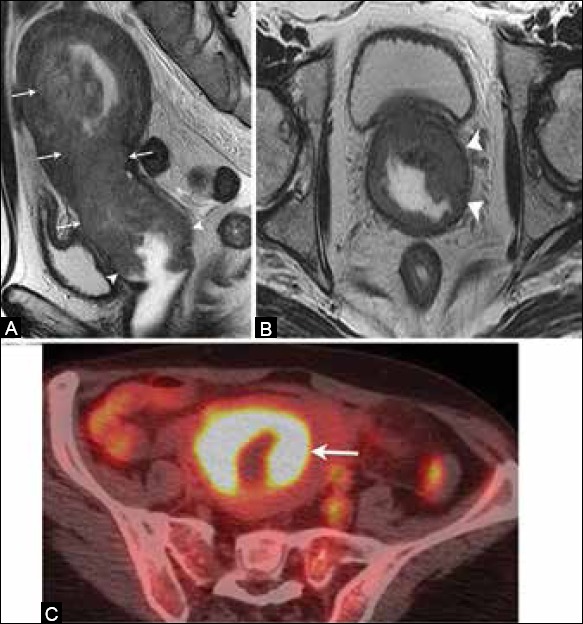
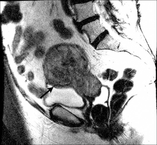
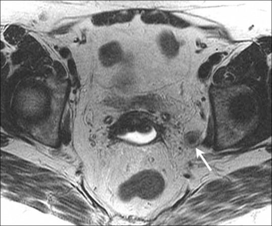
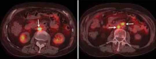
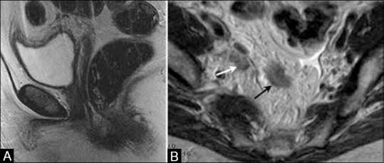
Endometrial carcinoma (EC) is the most common gynecologic malignancy in the United States. Prognosis depends on patient age, histological grade, depth of myometrial invasion and/or cervical invasion, and the presence of lymph node metastases. Although EC is staged surgically according to the International Federation of Gynecology and Obstetrics (FIGO) system, preoperative imaging can assist in optimal treatment planning. Several imaging techniques such as transvaginal ultrasonography (TVUS), computed tomography (CT), and magnetic resonance imaging (MRI) have been used as diagnostic tools for preoperative staging of EC. Recently, positron emission tomography (PET), PET/CT, and PET/MRI have also been used in staging these patients. In this article, we review the value of imaging in diagnosis, staging, treatment planning, and detection of recurrent disease in patients with EC.
子宫内膜癌(EC)是美国最常见的妇科恶性肿瘤。预后取决于患者年龄、组织学分级、肌层浸润深度和/或宫颈浸润情况以及淋巴结转移情况。尽管EC根据国际妇产科联合会(FIGO)系统进行手术分期,但术前影像学检查有助于制定最佳治疗方案。几种成像技术,如经阴道超声检查(TVUS)、计算机断层扫描(CT)和磁共振成像(MRI)已被用作EC术前分期的诊断工具。最近,正电子发射断层扫描(PET)、PET/CT和PET/MRI也被用于这些患者的分期。在本文中,我们综述了影像学在EC患者的诊断、分期、治疗方案制定及复发性疾病检测中的价值。