Chistiakov Dimitry A, Bobryshev Yuri V, Orekhov Alexander N
Department of Medical Nanobiotechnology, Pirogov Russian State Medical University, Moscow, Russia.
Faculty of Medicine and St Vincent's Centre for Applied Medical Research, University of New South Wales, Sydney, NSW, Australia.
J Cell Mol Med. 2015 Jun;19(6):1163-73. doi: 10.1111/jcmm.12591. Epub 2015 May 13.
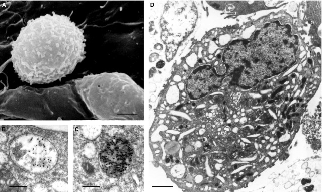
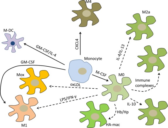
Macrophages display significant phenotypic heterogeneity. Two growth factors, macrophage colony-stimulating factor and chemokine (C-X-C motif) ligand 4, drive terminal differentiation of monocytes to M0 and M4 macrophages respectively. Compared to M0 macrophages, M4 cells have a unique transcriptome, with expression of surface markers such as S100A8, mannose receptor CD206 and matrix metalloproteinase 7. M4 macrophages did not express CD163, a scavenger receptor for haemoglobin/haptoglobin complex. Depending on the stimuli, M0 macrophages could polarize towards the proinflammatory M1 subset by treatment with lipopolysaccharide or interferon-γ. These macrophages produce a range of proinflammatory cytokines, nitric oxide, reactive oxygen species and exhibit high chemotactic and phagocytic activity. The alternative M2 type could be induced from M0 macrophage by stimulation with interleukin (IL)-4. M2 macrophages express high levels of CD206 and produce anti-inflammatory cytokines IL-10 and transforming growth factor-β. M1, M2 and M4 macrophages could be found in atherosclerotic plaques. In the plaque, macrophages are subjected to the intensive influence not only by cytokines and chemokines but also with bioactive lipids such as cholesterol and oxidized phospholipids. Oxidized phospholipids induce a distinct Mox phenotype in murine macrophages that express a unique panel of antioxidant enzymes under control of the redox-regulated transcription factor Klf2, resistant to lipid accumulation. In unstable human lesions, atheroprotective M(Hb) and HA-mac macrophage subsets could be found. These two subsets are induced by the haemoglobin/haptoglobin complex, highly express haeme oxygenase 1 and CD163, and are implicated in clearance of haemoglobin and erythrocyte remnants. In atherogenesis, the macrophage phenotype is plastic and could therefore be switched to proinflammatory (i.e. proatherogenic) and anti-inflammatory (i.e. atheroprotective). The aim of this review was to characterize changes in macrophage transcriptome in atherosclerosis and discuss key markers that characterize different phenotypes of macrophages present in atherosclerotic lesions.
巨噬细胞表现出显著的表型异质性。两种生长因子,即巨噬细胞集落刺激因子和趋化因子(C-X-C基序)配体4,分别驱动单核细胞向M0和M4巨噬细胞的终末分化。与M0巨噬细胞相比,M4细胞具有独特的转录组,表达诸如S100A8、甘露糖受体CD206和基质金属蛋白酶7等表面标志物。M4巨噬细胞不表达CD163,后者是血红蛋白/触珠蛋白复合物的清道夫受体。根据刺激因素的不同,M0巨噬细胞可通过脂多糖或干扰素-γ处理而极化为促炎性M1亚群。这些巨噬细胞产生一系列促炎细胞因子、一氧化氮、活性氧,并表现出高趋化性和吞噬活性。替代性M2型可通过白细胞介素(IL)-4刺激从M0巨噬细胞诱导产生。M2巨噬细胞高水平表达CD206,并产生抗炎细胞因子IL-10和转化生长因子-β。M1、M2和M4巨噬细胞均可在动脉粥样硬化斑块中发现。在斑块中,巨噬细胞不仅受到细胞因子和趋化因子的强烈影响,还受到生物活性脂质如胆固醇和氧化磷脂的影响。氧化磷脂在小鼠巨噬细胞中诱导出一种独特的Mox表型,该表型在氧化还原调节转录因子Klf2的控制下表达一组独特的抗氧化酶,对脂质积累具有抗性。在不稳定的人类病变中,可发现具有抗动脉粥样硬化作用的M(Hb)和HA-mac巨噬细胞亚群。这两个亚群由血红蛋白/触珠蛋白复合物诱导产生,高表达血红素加氧酶1和CD163,并参与血红蛋白和红细胞残余物的清除。在动脉粥样硬化发生过程中,巨噬细胞表型具有可塑性,因此可转变为促炎性(即促动脉粥样硬化性)和抗炎性(即抗动脉粥样硬化性)。本综述的目的是描述动脉粥样硬化中巨噬细胞转录组的变化,并讨论表征动脉粥样硬化病变中存在的不同巨噬细胞表型的关键标志物。