• 文献检索
  • 文档翻译
  • 深度研究
  • 学术资讯
  • Suppr Zotero 插件Zotero 插件
  • 邀请有礼
  • 套餐&价格
  • 历史记录
应用&插件
Suppr Zotero 插件Zotero 插件浏览器插件Mac 客户端Windows 客户端微信小程序
定价
高级版会员购买积分包购买API积分包
服务
文献检索文档翻译深度研究API 文档MCP 服务
关于我们
关于 Suppr公司介绍联系我们用户协议隐私条款
关注我们

Suppr 超能文献

核心技术专利:CN118964589B侵权必究
粤ICP备2023148730 号-1Suppr @ 2026

文献检索

告别复杂PubMed语法,用中文像聊天一样搜索,搜遍4000万医学文献。AI智能推荐,让科研检索更轻松。

立即免费搜索

文件翻译

保留排版,准确专业,支持PDF/Word/PPT等文件格式,支持 12+语言互译。

免费翻译文档

深度研究

AI帮你快速写综述,25分钟生成高质量综述,智能提取关键信息,辅助科研写作。

立即免费体验

几何细胞力学模型及从单细胞到有图案单层组织形成的细胞动力学拓扑算法

Mechanical model of geometric cell and topological algorithm for cell dynamics from single-cell to formation of monolayered tissues with pattern.

作者信息

Kachalo Sëma, Naveed Hammad, Cao Youfang, Zhao Jieling, Liang Jie

机构信息

Department of Bioengineering, The University of Illinois at Chicago, Chicago, IL, 60607.

Department of Bioengineering, The University of Illinois at Chicago, Chicago, IL, 60607; Computer, Electrical and Mathematical Sciences and Engineering Division, King Abdullah University of Science and Technology, Thuwal, Saudi Arabia.

出版信息

PLoS One. 2015 May 14;10(5):e0126484. doi: 10.1371/journal.pone.0126484. eCollection 2015.

DOI:10.1371/journal.pone.0126484
PMID:25974182
原文链接:https://pmc.ncbi.nlm.nih.gov/articles/PMC4431879/
Abstract

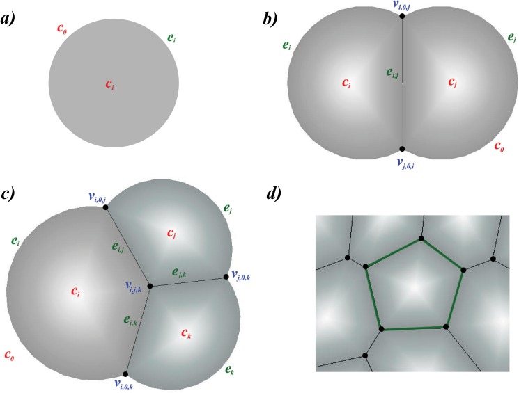
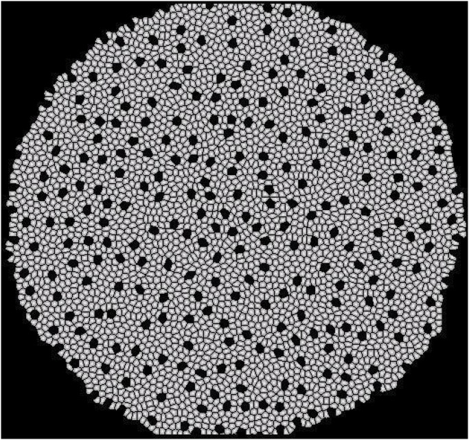
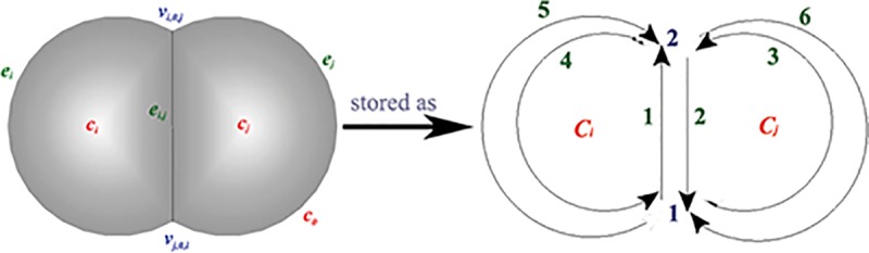
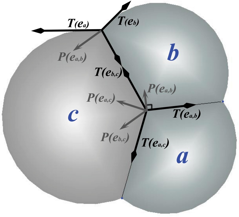
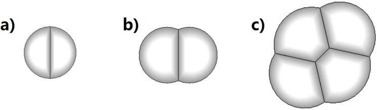
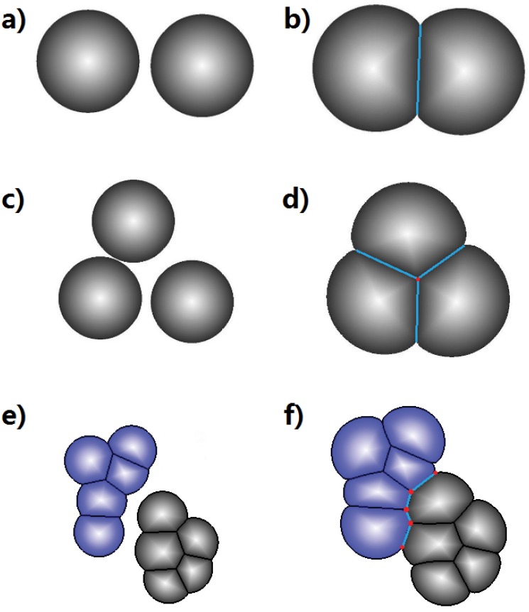
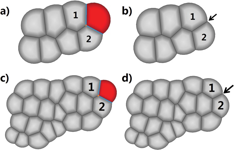
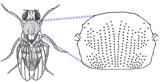
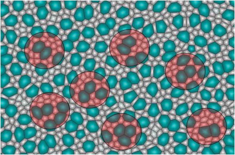
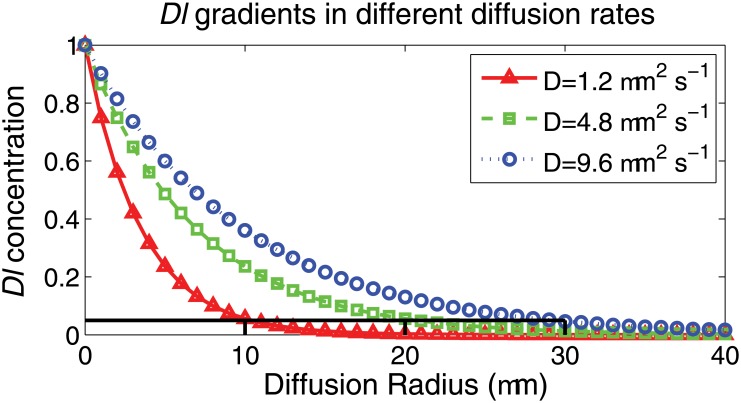
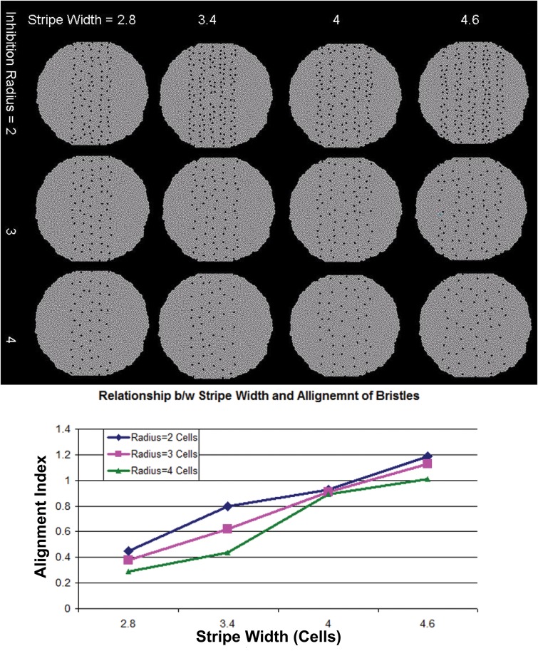
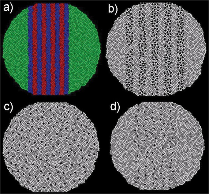
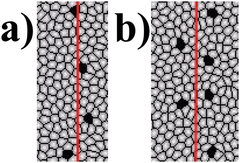
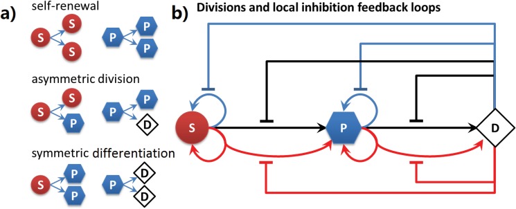
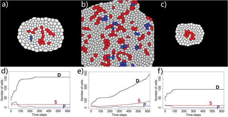
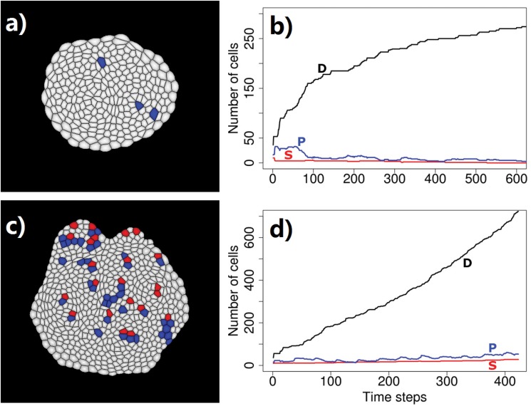
Geometric and mechanical properties of individual cells and interactions among neighboring cells are the basis of formation of tissue patterns. Understanding the complex interplay of cells is essential for gaining insight into embryogenesis, tissue development, and other emerging behavior. Here we describe a cell model and an efficient geometric algorithm for studying the dynamic process of tissue formation in 2D (e.g. epithelial tissues). Our approach improves upon previous methods by incorporating properties of individual cells as well as detailed description of the dynamic growth process, with all topological changes accounted for. Cell size, shape, and division plane orientation are modeled realistically. In addition, cell birth, cell growth, cell shrinkage, cell death, cell division, cell collision, and cell rearrangements are now fully accounted for. Different models of cell-cell interactions, such as lateral inhibition during the process of growth, can be studied in detail. Cellular pattern formation for monolayered tissues from arbitrary initial conditions, including that of a single cell, can also be studied in detail. Computational efficiency is achieved through the employment of a special data structure that ensures access to neighboring cells in constant time, without additional space requirement. We have successfully generated tissues consisting of more than 20,000 cells starting from 2 cells within 1 hour. We show that our model can be used to study embryogenesis, tissue fusion, and cell apoptosis. We give detailed study of the classical developmental process of bristle formation on the epidermis of D. melanogaster and the fundamental problem of homeostatic size control in epithelial tissues. Simulation results reveal significant roles of solubility of secreted factors in both the bristle formation and the homeostatic control of tissue size. Our method can be used to study broad problems in monolayered tissue formation. Our software is publicly available.

摘要

单个细胞的几何和力学特性以及相邻细胞之间的相互作用是组织模式形成的基础。理解细胞间复杂的相互作用对于深入了解胚胎发育、组织发育及其他新出现的行为至关重要。在此,我们描述一种细胞模型和一种高效的几何算法,用于研究二维(如上皮组织)中组织形成的动态过程。我们的方法通过纳入单个细胞的特性以及对动态生长过程的详细描述改进了先前的方法,同时考虑了所有拓扑变化。细胞大小、形状和分裂平面方向都得到了真实的建模。此外,现在还全面考虑了细胞出生、细胞生长、细胞收缩、细胞死亡、细胞分裂、细胞碰撞和细胞重排。可以详细研究不同的细胞 - 细胞相互作用模型,例如生长过程中的侧向抑制。从任意初始条件(包括单个细胞的初始条件)开始的单层组织的细胞模式形成也可以详细研究。通过采用一种特殊的数据结构实现了计算效率,该数据结构确保在固定时间内访问相邻细胞,且无需额外的空间要求。我们已成功在1小时内从2个细胞生成了由超过20,000个细胞组成的组织。我们表明我们的模型可用于研究胚胎发育、组织融合和细胞凋亡。我们详细研究了黑腹果蝇表皮上刚毛形成的经典发育过程以及上皮组织中稳态大小控制的基本问题。模拟结果揭示了分泌因子的溶解性在刚毛形成和组织大小的稳态控制中的重要作用。我们的方法可用于研究单层组织形成中的广泛问题。我们的软件可公开获取。

https://cdn.ncbi.nlm.nih.gov/pmc/blobs/bd5a/4431879/f90924b9a44f/pone.0126484.g020.jpg
https://cdn.ncbi.nlm.nih.gov/pmc/blobs/bd5a/4431879/4806d276e2a8/pone.0126484.g001.jpg
https://cdn.ncbi.nlm.nih.gov/pmc/blobs/bd5a/4431879/d9049a26ad4f/pone.0126484.g002.jpg
https://cdn.ncbi.nlm.nih.gov/pmc/blobs/bd5a/4431879/161d4f0b1c84/pone.0126484.g003.jpg
https://cdn.ncbi.nlm.nih.gov/pmc/blobs/bd5a/4431879/4357afd73376/pone.0126484.g004.jpg
https://cdn.ncbi.nlm.nih.gov/pmc/blobs/bd5a/4431879/bb18c855e58a/pone.0126484.g005.jpg
https://cdn.ncbi.nlm.nih.gov/pmc/blobs/bd5a/4431879/c5e4a90c2bd7/pone.0126484.g006.jpg
https://cdn.ncbi.nlm.nih.gov/pmc/blobs/bd5a/4431879/2684af04a17e/pone.0126484.g007.jpg
https://cdn.ncbi.nlm.nih.gov/pmc/blobs/bd5a/4431879/f2b7ffd7d313/pone.0126484.g008.jpg
https://cdn.ncbi.nlm.nih.gov/pmc/blobs/bd5a/4431879/fb3afe7fefd0/pone.0126484.g009.jpg
https://cdn.ncbi.nlm.nih.gov/pmc/blobs/bd5a/4431879/61386c8b0424/pone.0126484.g010.jpg
https://cdn.ncbi.nlm.nih.gov/pmc/blobs/bd5a/4431879/eeec63ea062b/pone.0126484.g011.jpg
https://cdn.ncbi.nlm.nih.gov/pmc/blobs/bd5a/4431879/4ccbdf02bbb3/pone.0126484.g012.jpg
https://cdn.ncbi.nlm.nih.gov/pmc/blobs/bd5a/4431879/f7c701699845/pone.0126484.g013.jpg
https://cdn.ncbi.nlm.nih.gov/pmc/blobs/bd5a/4431879/8b7564318e49/pone.0126484.g014.jpg
https://cdn.ncbi.nlm.nih.gov/pmc/blobs/bd5a/4431879/ccf0d9ae8fba/pone.0126484.g015.jpg
https://cdn.ncbi.nlm.nih.gov/pmc/blobs/bd5a/4431879/1cb5e4984e1d/pone.0126484.g016.jpg
https://cdn.ncbi.nlm.nih.gov/pmc/blobs/bd5a/4431879/93fe40023dd0/pone.0126484.g017.jpg
https://cdn.ncbi.nlm.nih.gov/pmc/blobs/bd5a/4431879/3a3b3da3d483/pone.0126484.g018.jpg
https://cdn.ncbi.nlm.nih.gov/pmc/blobs/bd5a/4431879/8435a4bc77d9/pone.0126484.g019.jpg
https://cdn.ncbi.nlm.nih.gov/pmc/blobs/bd5a/4431879/f90924b9a44f/pone.0126484.g020.jpg

相似文献

1
Mechanical model of geometric cell and topological algorithm for cell dynamics from single-cell to formation of monolayered tissues with pattern.几何细胞力学模型及从单细胞到有图案单层组织形成的细胞动力学拓扑算法
PLoS One. 2015 May 14;10(5):e0126484. doi: 10.1371/journal.pone.0126484. eCollection 2015.
2
Dynamic mechanical finite element model of biological cells for studying cellular pattern formation.用于研究细胞模式形成的生物细胞动态力学有限元模型。
Annu Int Conf IEEE Eng Med Biol Soc. 2013;2013:4517-20. doi: 10.1109/EMBC.2013.6610551.
3
Geometric order in proliferating epithelia: impact of rearrangements and cleavage plane orientation.增殖上皮中的几何秩序:重排和分裂平面取向的影响
Annu Int Conf IEEE Eng Med Biol Soc. 2010;2010:3808-11. doi: 10.1109/IEMBS.2010.5627601.
4
Apoptotic force and tissue dynamics during Drosophila embryogenesis.果蝇胚胎发育过程中的凋亡力与组织动力学
Science. 2008 Sep 19;321(5896):1683-6. doi: 10.1126/science.1157052.
5
Cell-Size Pleomorphism Drives Aberrant Clone Dispersal in Proliferating Epithelia.细胞大小多形性驱动增殖上皮中异常克隆的扩散。
Dev Cell. 2019 Oct 7;51(1):49-61.e4. doi: 10.1016/j.devcel.2019.08.005. Epub 2019 Sep 5.
6
Epithelial morphogenesis: apoptotic forces drive cell shape changes.上皮形态发生:凋亡力驱动细胞形状变化。
Dev Cell. 2015 Mar 9;32(5):532-3. doi: 10.1016/j.devcel.2015.02.020.
7
Developmental biology. Apoptosis turbocharges epithelial morphogenesis.发育生物学。细胞凋亡加速上皮形态发生。
Science. 2008 Sep 19;321(5896):1641-2. doi: 10.1126/science.1164583.
8
Modeling and inferring cleavage patterns in proliferating epithelia.增殖上皮细胞中分裂模式的建模与推断
PLoS Comput Biol. 2009 Jun;5(6):e1000412. doi: 10.1371/journal.pcbi.1000412. Epub 2009 Jun 12.
9
Mechanisms of regulating cell topology in proliferating epithelia: impact of division plane, mechanical forces, and cell memory.增殖上皮细胞中细胞拓扑结构调节的机制:分裂面、机械力和细胞记忆的影响。
PLoS One. 2012;7(8):e43108. doi: 10.1371/journal.pone.0043108. Epub 2012 Aug 17.
10
Cell topology, geometry, and morphogenesis in proliferating epithelia.增殖上皮中的细胞拓扑、几何形状和形态发生。
Curr Top Dev Biol. 2009;89:87-114. doi: 10.1016/S0070-2153(09)89004-2.

引用本文的文献

1
Evolutionary dynamics of mutants that modify population structure.改变种群结构的突变体的进化动态。
J R Soc Interface. 2023 Nov;20(208):20230355. doi: 10.1098/rsif.2023.0355. Epub 2023 Nov 29.
2
Free and Interfacial Boundaries in Individual-Based Models of Multicellular Biological systems.基于个体的多细胞生物系统模型中的自由和界面边界。
Bull Math Biol. 2023 Oct 8;85(11):111. doi: 10.1007/s11538-023-01214-8.
3
Recent Advances on the Model, Measurement Technique, and Application of Single Cell Mechanics.单细胞力学模型、测量技术及应用的最新进展。

本文引用的文献

1
Sequential emergence of the evenly spaced microchaetes on the notum of Drosophila.果蝇背板上均匀分布的微刚毛依次出现。
Rouxs Arch Dev Biol. 1993 Dec;203(3):151-158. doi: 10.1007/BF00365054.
2
Mechanisms of regulating tissue elongation in Drosophila wing: impact of oriented cell divisions, oriented mechanical forces, and reduced cell size.果蝇翅膀组织伸长的调控机制:定向细胞分裂、定向机械力和细胞尺寸减小的影响
PLoS One. 2014 Feb 4;9(2):e86725. doi: 10.1371/journal.pone.0086725. eCollection 2014.
3
Membrane tethered delta activates notch and reveals a role for spatio-mechanical regulation of the signaling pathway.
Int J Mol Sci. 2020 Aug 28;21(17):6248. doi: 10.3390/ijms21176248.
4
Cell-substrate mechanics guide collective cell migration through intercellular adhesion: a dynamic finite element cellular model.细胞-基质力学通过细胞间黏附指导细胞集体迁移:一个动态有限元细胞模型。
Biomech Model Mechanobiol. 2020 Oct;19(5):1781-1796. doi: 10.1007/s10237-020-01308-5. Epub 2020 Feb 27.
5
Dynamic cellular finite-element method for modelling large-scale cell migration and proliferation under the control of mechanical and biochemical cues: a study of re-epithelialization.用于在机械和生化信号控制下模拟大规模细胞迁移和增殖的动态细胞有限元方法:上皮再形成研究
J R Soc Interface. 2017 Apr;14(129). doi: 10.1098/rsif.2016.0959.
6
Multiscale Modeling of Cellular Epigenetic States: Stochasticity in Molecular Networks, Chromatin Folding in Cell Nuclei, and Tissue Pattern Formation of Cells.细胞表观遗传状态的多尺度建模:分子网络中的随机性、细胞核中的染色质折叠以及细胞的组织模式形成
Crit Rev Biomed Eng. 2015;43(4):323-46. doi: 10.1615/CritRevBiomedEng.2016016559.
7
TissueMiner: A multiscale analysis toolkit to quantify how cellular processes create tissue dynamics.组织挖掘器:一种多尺度分析工具包,用于量化细胞过程如何产生组织动态变化。
Elife. 2016 May 26;5:e14334. doi: 10.7554/eLife.14334.
膜结合的 Delta 激活 Notch,并揭示了信号通路的空间 - 机械调节的作用。
Biophys J. 2013 Dec 17;105(12):2655-65. doi: 10.1016/j.bpj.2013.11.012.
4
Modeling spatial population dynamics of stem cell lineage in wound healing and cancerogenesis.模拟伤口愈合和癌症发生过程中干细胞谱系的空间种群动态。
Annu Int Conf IEEE Eng Med Biol Soc. 2013;2013:5550-3. doi: 10.1109/EMBC.2013.6610807.
5
Modeling spatial population dynamics of stem cell lineage in tissue growth.组织生长过程中干细胞谱系的空间种群动态建模。
Annu Int Conf IEEE Eng Med Biol Soc. 2012;2012:5502-5. doi: 10.1109/EMBC.2012.6347240.
6
A model of epithelial invagination driven by collective mechanics of identical cells.由相同细胞的集体力学驱动的上皮内陷模型。
Biophys J. 2012 Sep 5;103(5):1069-77. doi: 10.1016/j.bpj.2012.07.018.
7
Mechanisms of regulating cell topology in proliferating epithelia: impact of division plane, mechanical forces, and cell memory.增殖上皮细胞中细胞拓扑结构调节的机制:分裂面、机械力和细胞记忆的影响。
PLoS One. 2012;7(8):e43108. doi: 10.1371/journal.pone.0043108. Epub 2012 Aug 17.
8
Mechanisms of tissue fusion during development.发育过程中组织融合的机制。
Development. 2012 May;139(10):1701-11. doi: 10.1242/dev.068338.
9
Free extracellular diffusion creates the Dpp morphogen gradient of the Drosophila wing disc.游离细胞外扩散作用创造了果蝇翅膀外胚层盘的 Dpp 形态发生梯度。
Curr Biol. 2012 Apr 24;22(8):668-75. doi: 10.1016/j.cub.2012.02.065. Epub 2012 Mar 22.
10
Mechanical forces mediate localized topological change in epithelia.机械力介导上皮组织中的局部拓扑变化。
Annu Int Conf IEEE Eng Med Biol Soc. 2011;2011:178-81. doi: 10.1109/IEMBS.2011.6089923.