• 文献检索
  • 文档翻译
  • 深度研究
  • 学术资讯
  • Suppr Zotero 插件Zotero 插件
  • 邀请有礼
  • 套餐&价格
  • 历史记录
应用&插件
Suppr Zotero 插件Zotero 插件浏览器插件Mac 客户端Windows 客户端微信小程序
定价
高级版会员购买积分包购买API积分包
服务
文献检索文档翻译深度研究API 文档MCP 服务
关于我们
关于 Suppr公司介绍联系我们用户协议隐私条款
关注我们

Suppr 超能文献

核心技术专利:CN118964589B侵权必究
粤ICP备2023148730 号-1Suppr @ 2026

文献检索

告别复杂PubMed语法,用中文像聊天一样搜索,搜遍4000万医学文献。AI智能推荐,让科研检索更轻松。

立即免费搜索

文件翻译

保留排版,准确专业,支持PDF/Word/PPT等文件格式,支持 12+语言互译。

免费翻译文档

深度研究

AI帮你快速写综述,25分钟生成高质量综述,智能提取关键信息,辅助科研写作。

立即免费体验

颅底磁共振成像的进展

Advances in magnetic resonance imaging of the skull base.

作者信息

Kirsch Claudia F E

机构信息

Department of Radiology, Wexner Medical Center, Ohio State University College of Medicine, Columbus, Ohio, United States.

出版信息

Int Arch Otorhinolaryngol. 2014 Oct;18(Suppl 2):S127-35. doi: 10.1055/s-0034-1390013.

DOI:10.1055/s-0034-1390013
PMID:25992137
原文链接:https://pmc.ncbi.nlm.nih.gov/articles/PMC4399586/
Abstract

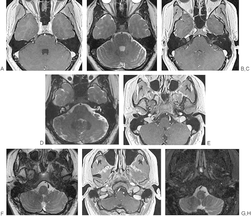
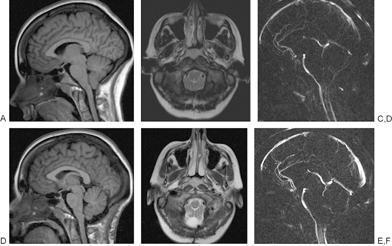
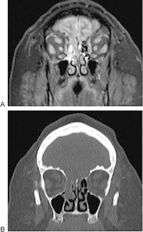
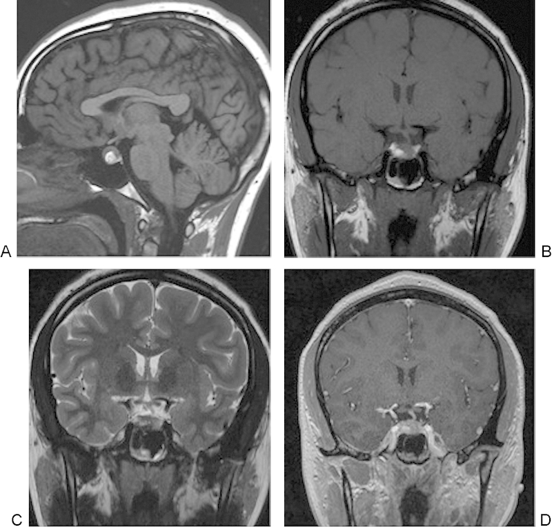
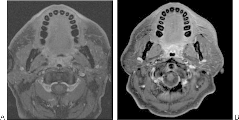
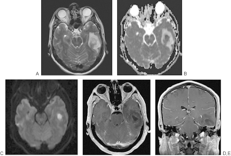
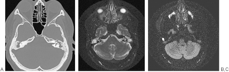
Introduction Over the past 20 years, magnetic resonance imaging (MRI) has advanced due to new techniques involving increased magnetic field strength and developments in coils and pulse sequences. These advances allow increased opportunity to delineate the complex skull base anatomy and may guide the diagnosis and treatment of the myriad of pathologies that can affect the skull base. Objectives The objective of this article is to provide a brief background of the development of MRI and illustrate advances in skull base imaging, including techniques that allow improved conspicuity, characterization, and correlative physiologic assessment of skull base pathologies. Data Synthesis Specific radiographic illustrations of increased skull base conspicuity including the lower cranial nerves, vessels, foramina, cerebrospinal fluid (CSF) leaks, and effacement of endolymph are provided. In addition, MRIs demonstrating characterization of skull base lesions, such as recurrent cholesteatoma versus granulation tissue or abscess versus tumor, are also provided as well as correlative clinical findings in CSF flow studies in a patient pre- and post-suboccipital decompression for a Chiari I malformation. Conclusions This article illustrates MRI radiographic advances over the past 20 years, which have improved clinicians' ability to diagnose, define, and hopefully improve the treatment and outcomes of patients with underlying skull base pathologies.

摘要

引言 在过去20年中,磁共振成像(MRI)因涉及更高磁场强度的新技术以及线圈和脉冲序列的发展而取得了进步。这些进展为描绘复杂的颅底解剖结构提供了更多机会,并可能指导对多种可影响颅底的病理状况的诊断和治疗。目的 本文的目的是提供MRI发展的简要背景,并阐述颅底成像的进展,包括能提高颅底病变的清晰度、特征描述及相关生理评估的技术。数据综合 提供了颅底清晰度提高的具体影像学例证,包括较低的颅神经、血管、孔道、脑脊液(CSF)漏以及内淋巴的消失。此外,还提供了显示颅底病变特征的MRI,如复发性胆脂瘤与肉芽组织或脓肿与肿瘤的鉴别,以及在一名Chiari I畸形患者枕下减压术前和术后进行的CSF流动研究中的相关临床发现。结论 本文阐述了过去20年中MRI影像学的进展,这些进展提高了临床医生诊断、界定并有望改善患有潜在颅底病变患者的治疗及预后的能力。

https://cdn.ncbi.nlm.nih.gov/pmc/blobs/feba/4399586/f4f78750e68a/10-1055-s-0034-1390013-i18s2a3ra-9.jpg
https://cdn.ncbi.nlm.nih.gov/pmc/blobs/feba/4399586/69453374463c/10-1055-s-0034-1390013-i18s2a3ra-1.jpg
https://cdn.ncbi.nlm.nih.gov/pmc/blobs/feba/4399586/472f35f2e9b4/10-1055-s-0034-1390013-i18s2a3ra-2.jpg
https://cdn.ncbi.nlm.nih.gov/pmc/blobs/feba/4399586/63ad242e2564/10-1055-s-0034-1390013-i18s2a3ra-3.jpg
https://cdn.ncbi.nlm.nih.gov/pmc/blobs/feba/4399586/1e3ba8a7bb09/10-1055-s-0034-1390013-i18s2a3ra-4.jpg
https://cdn.ncbi.nlm.nih.gov/pmc/blobs/feba/4399586/4c67f8179ea0/10-1055-s-0034-1390013-i18s2a3ra-5.jpg
https://cdn.ncbi.nlm.nih.gov/pmc/blobs/feba/4399586/8dd786850b46/10-1055-s-0034-1390013-i18s2a3ra-6.jpg
https://cdn.ncbi.nlm.nih.gov/pmc/blobs/feba/4399586/ade0481e8dae/10-1055-s-0034-1390013-i18s2a3ra-7.jpg
https://cdn.ncbi.nlm.nih.gov/pmc/blobs/feba/4399586/1ee8c4077102/10-1055-s-0034-1390013-i18s2a3ra-8.jpg
https://cdn.ncbi.nlm.nih.gov/pmc/blobs/feba/4399586/f4f78750e68a/10-1055-s-0034-1390013-i18s2a3ra-9.jpg

相似文献

1
Advances in magnetic resonance imaging of the skull base.颅底磁共振成像的进展
Int Arch Otorhinolaryngol. 2014 Oct;18(Suppl 2):S127-35. doi: 10.1055/s-0034-1390013.
2
Radiographic enhancement of the nasoseptal flap does not predict postoperative cerebrospinal fluid leaks in endoscopic skull base reconstruction.鼻内隔瓣的影像学增强并不预示内镜颅底重建术后的脑脊液漏。
Laryngoscope. 2012 Jun;122(6):1226-34. doi: 10.1002/lary.23351. Epub 2012 May 7.
3
Special MRI Techniques to Suss out Spontaneous Cerebrospinal Fluid Leaks.特殊 MRI 技术探查自发性脑脊液漏。
Top Magn Reson Imaging. 2021 Jun 1;30(3):159-166. doi: 10.1097/RMR.0000000000000281.
4
Imaging of skull base lesions.颅底病变的影像学检查。
Handb Clin Neurol. 2016;135:637-657. doi: 10.1016/B978-0-444-53485-9.00030-1.
5
Magnetic resonance cisternogram with intrathecal gadolinium with delayed imaging for difficult to diagnose cerebrospinal fluid leaks of anterior skull base.磁共振脑池造影术,鞘内注射钆后延迟成像,用于诊断前颅底脑脊液漏困难的病例。
Int Forum Allergy Rhinol. 2015 Apr;5(4):333-8. doi: 10.1002/alr.21475. Epub 2015 Jan 5.
6
Imaging of Acquired Skull Base Cerebrospinal Fluid Leaks.后天性颅底脑脊液漏的影像学评估
Neuroimaging Clin N Am. 2021 Nov;31(4):509-522. doi: 10.1016/j.nic.2021.05.009.
7
Imaging of skull base pathologies: Role of advanced magnetic resonance imaging techniques.颅底病变的影像学检查:先进磁共振成像技术的作用
Neuroradiol J. 2015 Aug;28(4):426-37. doi: 10.1177/1971400915609341. Epub 2015 Oct 1.
8
Venous Sinus Stenting in the Management of Patients with Intracranial Hypertension Manifesting with Skull Base Cerebrospinal Fluid Leaks.静脉窦支架置入术治疗合并颅底脑脊液漏的颅内高压患者
World Neurosurg. 2017 Oct;106:103-112. doi: 10.1016/j.wneu.2017.06.087. Epub 2017 Jun 20.
9
Efficacy of the pedicled nasoseptal flap without cerebrospinal fluid (CSF) diversion for repair of skull base defects: incidence of postoperative CSF leaks.带蒂鼻中隔-鼻黏膜瓣修复颅底缺损而不进行脑脊液(CSF)转流的疗效:术后脑脊液漏的发生率。
Int Forum Allergy Rhinol. 2012 Sep-Oct;2(5):397-401. doi: 10.1002/alr.21040. Epub 2012 Apr 23.
10
[Anatomy of the skull base and the cranial nerves in slice imaging].[切片成像中颅底及脑神经的解剖学]
Radiologe. 2009 Jul;49(7):584-97. doi: 10.1007/s00117-008-1800-0.

引用本文的文献

1
Infra-Temporal and Pterygo-Palatine Fossae Tumors: A Frontier in Endoscopic Endonasal Surgery-Description of the Surgical Anatomy of the Approach and Report of Illustrative Cases.翼腭窝和颞下窝肿瘤:内镜经鼻颅底手术的前沿——手术入路解剖描述及典型病例报告。
Int J Environ Res Public Health. 2022 May 25;19(11):6413. doi: 10.3390/ijerph19116413.
2
Averting Delayed Complications of Open Anterior Skull Base Surgery.避免开放性前颅底手术的延迟并发症
J Neurol Surg B Skull Base. 2020 Aug 5;82(4):450-455. doi: 10.1055/s-0040-1714097. eCollection 2021 Aug.
3
Surgical management and follow-up of lateral skull base tumors: An 8-year review.

本文引用的文献

1
Diffusion-weighted MRI "claw sign" improves differentiation of infectious from degenerative modic type 1 signal changes of the spine.扩散加权磁共振成像“爪征”有助于鉴别脊柱感染性与退行性Modic 1型信号改变。
AJNR Am J Neuroradiol. 2014 Aug;35(8):1647-52. doi: 10.3174/ajnr.A3948. Epub 2014 Apr 17.
2
Experience with intraoperative navigation and imaging during endoscopic transnasal spinal approaches to the foramen magnum and odontoid.经鼻内镜下枕大孔和齿状突脊柱手术中术中导航和成像的经验。
Neurosurg Focus. 2014 Mar;36(3):E4. doi: 10.3171/2014.1.FOCUS13533.
3
Detection and grading of endolymphatic hydrops in Menière disease using MR imaging.
侧颅底肿瘤的外科治疗与随访:一项8年回顾
Mol Clin Oncol. 2017 Feb;6(2):214-220. doi: 10.3892/mco.2017.1126. Epub 2017 Jan 10.
4
The pterygopalatine fossa: imaging anatomy, communications, and pathology revisited.翼腭窝:影像学解剖、连通关系及病理学再探讨
Insights Imaging. 2016 Aug;7(4):589-99. doi: 10.1007/s13244-016-0498-1. Epub 2016 May 26.
利用磁共振成像检测梅尼埃病内淋巴积水并进行分级
AJNR Am J Neuroradiol. 2014 Jul;35(7):1387-92. doi: 10.3174/ajnr.A3856. Epub 2014 Feb 13.
4
Arterial, venous, and cerebrospinal fluid flow: simultaneous assessment with Bayesian multipoint velocity-encoded MR imaging.动脉、静脉和脑脊液流动:基于贝叶斯多点流速编码磁共振成像的同步评估。
Radiology. 2014 Feb;270(2):566-73. doi: 10.1148/radiol.13130840. Epub 2013 Oct 30.
5
Use of preoperative MRI to predict vestibular schwannoma intraoperative consistency and facial nerve outcome.使用术前磁共振成像预测前庭神经鞘瘤术中的质地及面神经预后。
J Neurol Surg B Skull Base. 2013 Dec;74(6):347-50. doi: 10.1055/s-0033-1347369. Epub 2013 May 22.
6
Analysis of cerebrospinal fluid flow dynamics and morphology in Chiari I malformation with cine phase-contrast magnetic resonance imaging.利用电影相位对比磁共振成像分析Chiari I型畸形中的脑脊液流动动力学和形态学。
Acta Neurochir (Wien). 2014 Apr;156(4):707-13. doi: 10.1007/s00701-013-1958-8. Epub 2014 Jan 7.
7
3T MRI and 128-slice dual-source CT cisternography images of the cranial nerves a brief pictorial review for clinicians.3T磁共振成像和128层双源CT脑池造影术对颅神经成像:给临床医生的简要图像综述
Clin Anat. 2014 Jan;27(1):31-45. doi: 10.1002/ca.22311. Epub 2013 Dec 2.
8
[Endolymphatic hydrops detected with inner ear gd contrast-enhanced MRI; comparison between administration routes or with ECochG or glycerol test].[内耳钆对比增强磁共振成像检测到的内淋巴积水;给药途径之间或与耳蜗电图或甘油试验的比较]
Nihon Jibiinkoka Gakkai Kaiho. 2013 Aug;116(8):960-8. doi: 10.3950/jibiinkoka.116.960.
9
Non-echo planar diffusion-weighted MRI increases follow-up accuracy after one-step step canal wall-down obliteration surgery for cholesteatoma.非回波平面扩散加权磁共振成像提高了胆脂瘤一步法外耳道壁下闭合手术后的随访准确性。
Acta Otolaryngol. 2013 Jun;133(6):574-83. doi: 10.3109/00016489.2012.762118.
10
Diffusion-weighted MRI: distinction of skull base chordoma from chondrosarcoma.弥散加权 MRI:颅底脊索瘤与软骨肉瘤的鉴别。
AJNR Am J Neuroradiol. 2013 May;34(5):1056-61, S1. doi: 10.3174/ajnr.A3333. Epub 2012 Nov 1.