Sun Zhenxing, Cheng Tsung O, Li Ling, Zhang Li, Wang Xinfang, Dong Nianguo, Lv Qing, Li Ke, Yuan Li, Wang Jing, Xie Mingxing
Department of Ultrasound, Union Hospital, Tongji Medical College, Huazhong University of Science and Technology, Hubei Province Key Laboratory of Molecular Imaging, Wuhan, People's Republic of China.
Department of Ultrasound, Union Hospital, Tongji Medical College, Huazhong University of Science and Technology, Hubei Province Key Laboratory of Molecular Imaging, Wuhan, People's Republic of China; Department of Medicine, George Washington University Medical Center, 2150 Pennsylvania Avenue N. W., Washington, D. C., United States of America.
PLoS One. 2015 Jun 1;10(6):e0127399. doi: 10.1371/journal.pone.0127399. eCollection 2015.
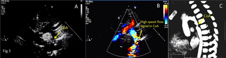
Although aortography is well known as the "gold standard" for the diagnosis of coarctation of aorta (CoA), the method is invasive, expensive and not readily accepted by some patients. Ultrasound diagnosis for CoA is non-invasive, inexpensive, readily accepted by every patient, and can be repeated as frequently as necessary. The purpose of this presentation is to evaluate the applicability of transthoracic echocardiography for the diagnosis of CoA. The echocardiographic appearances of 53 patients with CoA who had undergone surgery during a 5-year period from January 2008 to October 2012 were analyzed retrospectively, and the results were compared with findings at surgery. Fifty-three patients with CoA include six with isolated CoA and 47 of CoA associated with other cardiac anomalies. Of the 53 operated patients, 48 were correctly diagnosed preoperatively by echocardiography, while two were misdiagnosed as interrupted aortic arch and the diagnosis were missed in three other patients. Thus the diagnostic accuracy rate was 90.6%, and the misdiagnosis rate was 9.4%. Preoperative echocardiographic evaluation offers very satisfactory anatomic assessment in most patients with CoA. It makes preoperative angiography unnecessary. Thus transthoracic echocardiography should be the first-line method for the diagnosis of coarctation of the aorta.
尽管主动脉造影术作为诊断主动脉缩窄(CoA)的“金标准”广为人知,但该方法具有侵入性、费用高昂且一些患者不易接受。CoA的超声诊断具有非侵入性、成本低廉、易被每位患者接受且可根据需要频繁重复的特点。本报告的目的是评估经胸超声心动图在CoA诊断中的适用性。回顾性分析了2008年1月至2012年10月期间5年中接受手术的53例CoA患者的超声心动图表现,并将结果与手术所见进行比较。53例CoA患者中,6例为孤立性CoA,47例为合并其他心脏畸形的CoA。在53例接受手术的患者中,48例术前经超声心动图正确诊断,2例误诊为主动脉弓中断,另有3例漏诊。因此诊断准确率为90.6%,误诊率为9.4%。术前超声心动图评估在大多数CoA患者中提供了非常令人满意的解剖学评估。它使术前血管造影变得不必要。因此经胸超声心动图应作为主动脉缩窄诊断的一线方法。