List Jonathan, Ott Stefanie, Bukowski Martin, Lindenberg Robert, Flöel Agnes
Department of Neurology, Charité Universitätsmedizin Berlin Berlin, Germany.
Department of Neurology, Charité Universitätsmedizin Berlin Berlin, Germany ; Center for Stroke Research Berlin, Charité Universitätsmedizin Berlin Berlin, Germany.
Front Hum Neurosci. 2015 May 21;9:228. doi: 10.3389/fnhum.2015.00228. eCollection 2015.
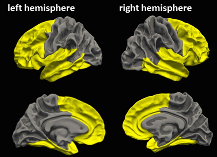
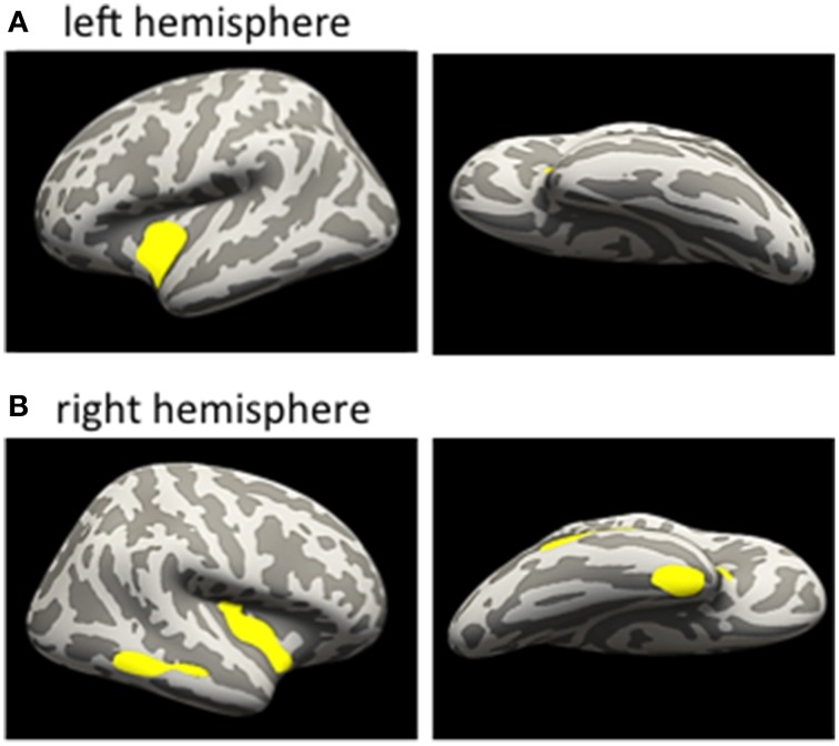
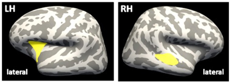
Recurrent mild traumatic brain injuries (mTBIs) are regarded as an independent risk factor for developing dementia in later life. We here aimed to evaluate associations between recurrent mTBIs, cognition, and gray matter volume and microstructure as revealed by structural magnetic resonance imaging (MRI) in the chronic phase after mTBIs in young adulthood. We enrolled 20 young-to-middle-aged subjects, who reported two or more sports-related mTBIs, with the last mTBI > 6 months prior to study enrolment (mTBI group), and 21 age-, sex- and education matched controls with no history of mTBI (control group). All participants received comprehensive neuropsychological testing, and high resolution T1-weighted and diffusion tensor MRI in order to assess cortical thickness (CT) and microstructure, hippocampal volume, and ventricle size. Compared to the control group, subjects of the mTBI group presented with lower CT within the right temporal lobe and left insula using an a priori region of interest approach. Higher number of mTBIs was associated with lower CT in bilateral insula, right middle temporal gyrus and right entorhinal area. Our results suggest persistent detrimental effects of recurrent mTBIs on CT already in young-to-middle-aged adults. If additional structural deterioration occurs during aging, subtle neuropsychological decline may progress to clinically overt dementia earlier than in age-matched controls, a hypothesis to be assessed in future prospective trials.
反复轻度创伤性脑损伤(mTBI)被视为晚年患痴呆症的独立危险因素。我们的目的是评估反复mTBI、认知与灰质体积和微观结构之间的关联,这些关联通过结构磁共振成像(MRI)在年轻成年人mTBI慢性期揭示。我们招募了20名年轻至中年受试者,他们报告有两次或更多与运动相关的mTBI,最后一次mTBI发生在研究入组前>6个月(mTBI组),以及21名年龄、性别和教育程度匹配且无mTBI病史的对照组(对照组)。所有参与者都接受了全面的神经心理学测试,以及高分辨率T1加权和扩散张量MRI,以评估皮质厚度(CT)和微观结构、海马体积和脑室大小。与对照组相比,mTBI组的受试者使用先验感兴趣区域方法,右侧颞叶和左侧岛叶的CT较低。mTBI次数越多,双侧岛叶、右侧颞中回和右侧内嗅区的CT越低。我们的结果表明,反复mTBI对年轻至中年成年人的CT已经存在持续的有害影响。如果在衰老过程中发生额外的结构恶化,轻微的神经心理学衰退可能比年龄匹配的对照组更早发展为临床明显的痴呆症,这一假设有待未来前瞻性试验评估。