• 文献检索
  • 文档翻译
  • 深度研究
  • 学术资讯
  • Suppr Zotero 插件Zotero 插件
  • 邀请有礼
  • 套餐&价格
  • 历史记录
应用&插件
Suppr Zotero 插件Zotero 插件浏览器插件Mac 客户端Windows 客户端微信小程序
定价
高级版会员购买积分包购买API积分包
服务
文献检索文档翻译深度研究API 文档MCP 服务
关于我们
关于 Suppr公司介绍联系我们用户协议隐私条款
关注我们

Suppr 超能文献

核心技术专利:CN118964589B侵权必究
粤ICP备2023148730 号-1Suppr @ 2026

文献检索

告别复杂PubMed语法,用中文像聊天一样搜索,搜遍4000万医学文献。AI智能推荐,让科研检索更轻松。

立即免费搜索

文件翻译

保留排版,准确专业,支持PDF/Word/PPT等文件格式,支持 12+语言互译。

免费翻译文档

深度研究

AI帮你快速写综述,25分钟生成高质量综述,智能提取关键信息,辅助科研写作。

立即免费体验

羊 Jamshidi 活检缺损中骨诱导性软骨诱导:软骨下壳聚糖-血植入物治疗和不治疗的 1 天、3 周和 3 个月修复。

Bone-Induced Chondroinduction in Sheep Jamshidi Biopsy Defects with and without Treatment by Subchondral Chitosan-Blood Implant: 1-Day, 3-Week, and 3-Month Repair.

机构信息

Department of Clinical Studies, University of Guelph, Guelph, Ontario, Canada.

Department of Chemical Engineering, École Polytechnique, Montreal, Quebec, Canada.

出版信息

Cartilage. 2013 Apr;4(2):131-43. doi: 10.1177/1947603512463227.

DOI:10.1177/1947603512463227
PMID:26069656
原文链接:https://pmc.ncbi.nlm.nih.gov/articles/PMC4297102/
Abstract

OBJECTIVE

Delivery of chitosan to subchondral bone is a novel approach for augmented marrow stimulation. We evaluated the effect of 3 presolidified chitosan-blood implant formulations on osteochondral repair progression compared with untreated defects.

DESIGN

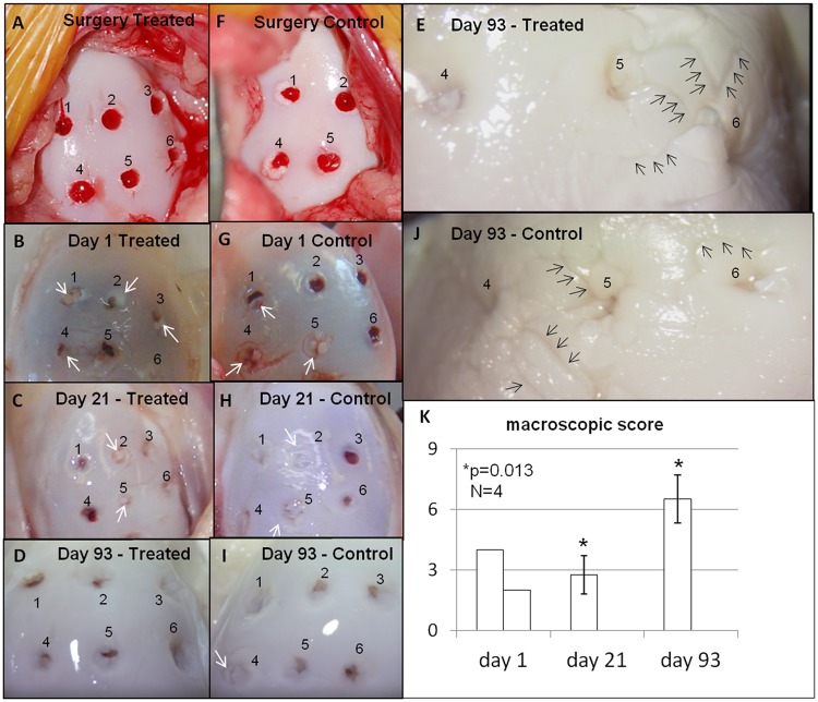
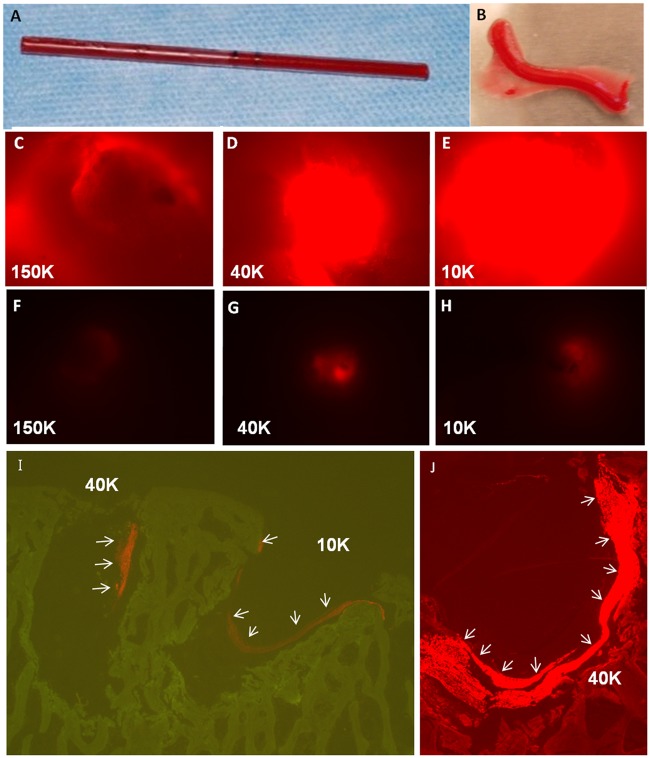
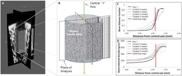
In N = 5 adult sheep, six 2-mm diameter Jamshidi biopsy holes were created bilaterally in the medial femoral condyle and treated with presolidified chitosan-blood implant with fluorescent chitosan tracer (10 kDa, 40 kDa, or 150k Da chitosan, left knee) or left to bleed (untreated, right knee). Implant residency and osteochondral repair were assessed at 1 day (N = 1), 3 weeks (N = 2), or 3 months (N = 2) postoperative using fluorescence microscopy, histomorphometry, stereology, and micro-computed tomography.

RESULTS

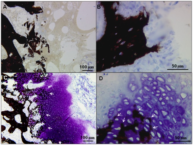
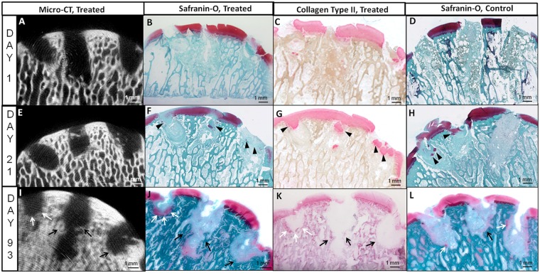
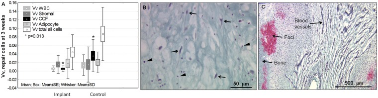
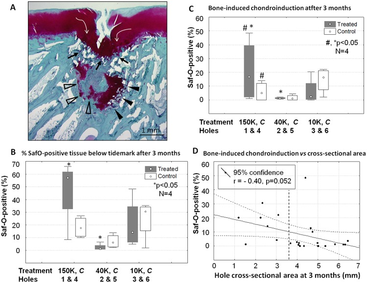
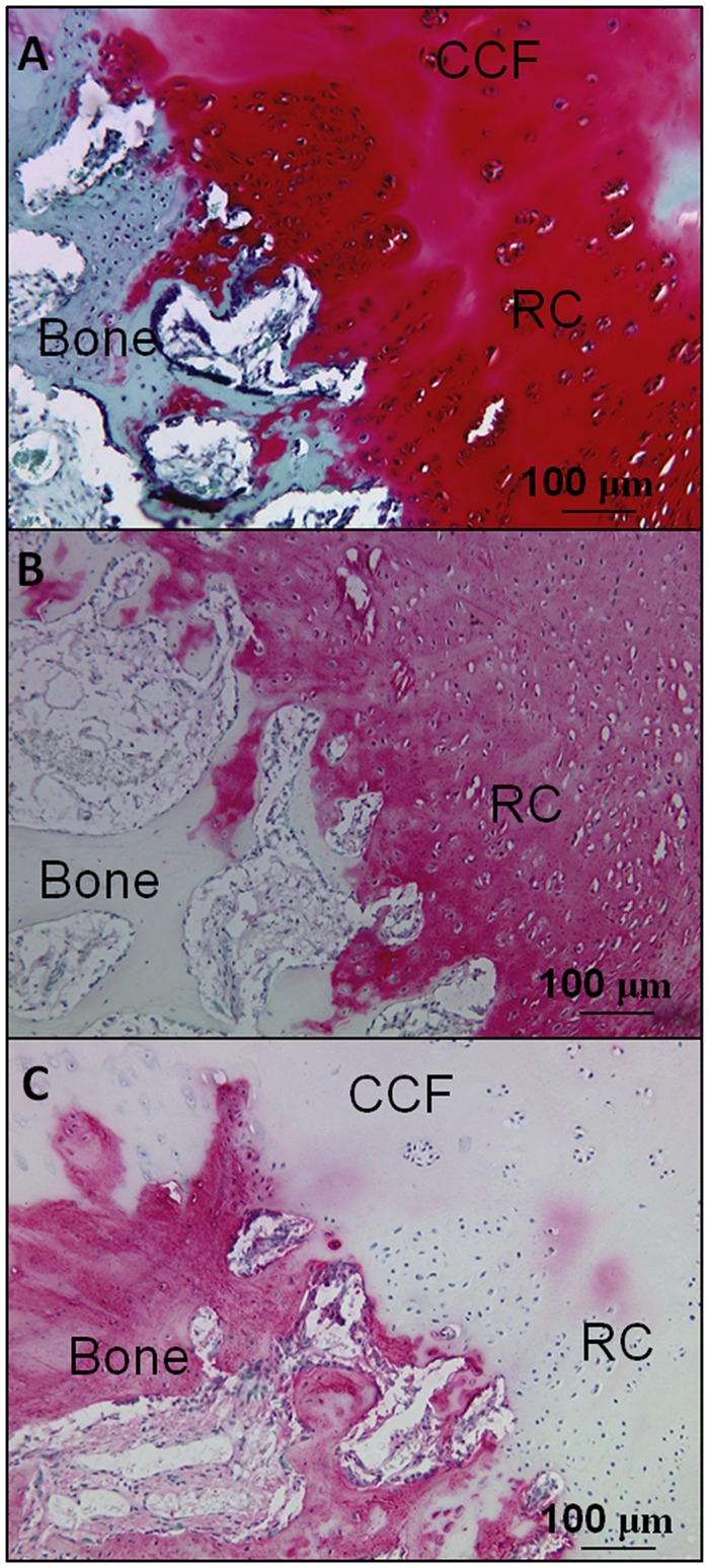
Chitosan implants were retained in 89% of treated Jamshidi holes up to 3 weeks postoperative. At 3 weeks, biopsy sites were variably covered by cartilage flow, and most bone holes contained cartilage flow fragments and heterogeneous granulation tissues with sparse leukocytes, stromal cells, and occasional adipocytes (volume density 1% to 3%). After 3 months of repair, most Jamshidi bone holes were deeper, remodeling at the edges, filled with angiogenic granulation tissue, and lined with variably sized chondrogenic foci fused to bone trabeculae or actively repairing bone plate. The 150-kDa chitosan implant elicited more subchondral cartilage formation compared with 40-kDa chitosan-treated and control defects (P < 0.05, N = 4). Treated defects contained more mineralized repair tissue than control defects at 3 months (P < 0.05, N = 12).

CONCLUSION

Bone plate-induced chondroinduction is an articular cartilage repair mechanism. Jamshidi biopsy repair takes longer than 3 months and can be influenced by subchondral chitosan-blood implant.

摘要

目的

壳聚糖递送至软骨下骨是增强骨髓刺激的一种新方法。我们评估了 3 种预固化壳聚糖-血植入物制剂与未处理的缺陷相比对骨软骨修复进展的影响。

设计

在 N = 5 只成年绵羊中,双侧股骨内侧髁共创建了 6 个 2-mm 直径的 Jamshidi 活检孔,并用预固化的壳聚糖-血植入物(10 kDa、40 kDa 或 150 kDa 壳聚糖,左膝)或让其出血(未处理,右膝)处理。术后 1 天(N = 1)、3 周(N = 2)或 3 个月(N = 2),使用荧光显微镜、组织形态计量学、体视学和微计算机断层扫描评估植入物的驻留和骨软骨修复情况。

结果

壳聚糖植入物在术后 3 周内保持在 89%的治疗性 Jamshidi 孔中。在 3 周时,活检部位被软骨流不同程度地覆盖,大多数骨孔含有软骨流碎片和异质肉芽组织,有稀疏的白细胞、基质细胞和偶尔的脂肪细胞(体积密度为 1%至 3%)。在 3 个月的修复后,大多数 Jamshidi 骨孔更深,边缘重塑,充满血管生成的肉芽组织,并有大小不一的软骨生成灶融合到骨小梁或积极修复骨板。与 40 kDa 壳聚糖处理的和对照缺陷相比,150 kDa 壳聚糖植入物引起更多的软骨下软骨形成(P < 0.05,N = 4)。在 3 个月时,治疗性缺陷的矿化修复组织比对照缺陷多(P < 0.05,N = 12)。

结论

骨板诱导的软骨诱导是一种关节软骨修复机制。Jamshidi 活检修复需要超过 3 个月的时间,并且可以受到软骨下壳聚糖-血植入物的影响。

https://cdn.ncbi.nlm.nih.gov/pmc/blobs/4820/4297102/13feb1e90f88/10.1177_1947603512463227-fig8.jpg
https://cdn.ncbi.nlm.nih.gov/pmc/blobs/4820/4297102/df793739a6d8/10.1177_1947603512463227-fig1.jpg
https://cdn.ncbi.nlm.nih.gov/pmc/blobs/4820/4297102/4f51dc02cbdc/10.1177_1947603512463227-fig2.jpg
https://cdn.ncbi.nlm.nih.gov/pmc/blobs/4820/4297102/460bc0d0b23b/10.1177_1947603512463227-fig3.jpg
https://cdn.ncbi.nlm.nih.gov/pmc/blobs/4820/4297102/d46f3f3694e5/10.1177_1947603512463227-fig4.jpg
https://cdn.ncbi.nlm.nih.gov/pmc/blobs/4820/4297102/054ec76aab72/10.1177_1947603512463227-fig5.jpg
https://cdn.ncbi.nlm.nih.gov/pmc/blobs/4820/4297102/9825fa1e16fb/10.1177_1947603512463227-fig6.jpg
https://cdn.ncbi.nlm.nih.gov/pmc/blobs/4820/4297102/5fefd83a90ce/10.1177_1947603512463227-fig7.jpg
https://cdn.ncbi.nlm.nih.gov/pmc/blobs/4820/4297102/13feb1e90f88/10.1177_1947603512463227-fig8.jpg

相似文献

1
Bone-Induced Chondroinduction in Sheep Jamshidi Biopsy Defects with and without Treatment by Subchondral Chitosan-Blood Implant: 1-Day, 3-Week, and 3-Month Repair.羊 Jamshidi 活检缺损中骨诱导性软骨诱导:软骨下壳聚糖-血植入物治疗和不治疗的 1 天、3 周和 3 个月修复。
Cartilage. 2013 Apr;4(2):131-43. doi: 10.1177/1947603512463227.
2
Effect of a Rapidly Degrading Presolidified 10 kDa Chitosan/Blood Implant and Subchondral Marrow Stimulation Surgical Approach on Cartilage Resurfacing in a Sheep Model.快速降解的预固化10 kDa壳聚糖/血液植入物及软骨下骨髓刺激手术方法对绵羊模型软骨修复的影响
Cartilage. 2017 Oct;8(4):417-431. doi: 10.1177/1947603516676872. Epub 2016 Nov 11.
3
Subchondral chitosan/blood implant-guided bone plate resorption and woven bone repair is coupled to hyaline cartilage regeneration from microdrill holes in aged rabbit knees.软骨下壳聚糖/血液植入物引导的骨板吸收和编织骨修复与老年兔膝关节微钻孔处的透明软骨再生相关联。
Osteoarthritis Cartilage. 2014 Feb;22(2):323-33. doi: 10.1016/j.joca.2013.12.011. Epub 2013 Dec 19.
4
Microdrilled cartilage defects treated with thrombin-solidified chitosan/blood implant regenerate a more hyaline, stable, and structurally integrated osteochondral unit compared to drilled controls.经凝血酶固化壳聚糖/血植入物治疗的微钻取软骨缺损与钻孔对照相比,再生出更透明、稳定且结构整合的软骨-骨单位。
Tissue Eng Part A. 2012 Mar;18(5-6):508-19. doi: 10.1089/ten.TEA.2011.0178. Epub 2011 Nov 11.
5
Stereological analysis of subchondral angiogenesis induced by chitosan and coagulation factors in microdrilled articular cartilage defects.微钻削关节软骨缺损中壳聚糖和凝血因子诱导的软骨下血管生成的体视学分析。
Osteoarthritis Cartilage. 2013 Jun;21(6):849-59. doi: 10.1016/j.joca.2013.03.012. Epub 2013 Mar 21.
6
Chitosan-glycerol phosphate/blood implants elicit hyaline cartilage repair integrated with porous subchondral bone in microdrilled rabbit defects.壳聚糖-甘油磷酸酯/血液植入物可引发透明软骨修复,并与微钻孔兔缺损处的多孔软骨下骨整合。
Osteoarthritis Cartilage. 2007 Jan;15(1):78-89. doi: 10.1016/j.joca.2006.06.015. Epub 2006 Aug 8.
7
Subchondral pre-solidified chitosan/blood implants elicit reproducible early osteochondral wound-repair responses including neutrophil and stromal cell chemotaxis, bone resorption and repair, enhanced repair tissue integration and delayed matrix deposition.骨下预凝固壳聚糖/血植入物可引发可重复的早期骨软骨创面修复反应,包括中性粒细胞和基质细胞趋化作用、骨质吸收和修复、增强修复组织整合以及延迟基质沉积。
BMC Musculoskelet Disord. 2013 Jan 16;14:27. doi: 10.1186/1471-2474-14-27.
8
Acute Osteoclast Activity following Subchondral Drilling Is Promoted by Chitosan and Associated with Improved Cartilage Repair Tissue Integration.软骨下钻孔后破骨细胞活性增强,壳聚糖促进其活性,有助于改善软骨修复组织的整合。
Cartilage. 2011 Apr;2(2):173-85. doi: 10.1177/1947603510381096.
9
Chitosan-glycerol phosphate/blood implants increase cell recruitment, transient vascularization and subchondral bone remodeling in drilled cartilage defects.壳聚糖-甘油磷酸酯/血液植入物可增加钻孔软骨缺损处的细胞募集、短暂血管化和软骨下骨重塑。
Osteoarthritis Cartilage. 2007 Mar;15(3):316-27. doi: 10.1016/j.joca.2006.08.007. Epub 2006 Sep 26.
10
Scaffold-guided subchondral bone repair: implication of neutrophils and alternatively activated arginase-1+ macrophages.支架引导的软骨下骨修复:中性粒细胞和替代性激活的精氨酸酶 1+巨噬细胞的作用。
Am J Sports Med. 2010 Sep;38(9):1845-56. doi: 10.1177/0363546510369547. Epub 2010 Jun 3.

引用本文的文献

1
CARGEL Bioscaffold improves cartilage repair tissue after bone marrow stimulation in a minipig model.CARGEL生物支架在小型猪模型中促进骨髓刺激后的软骨修复组织。
J Exp Orthop. 2020 May 8;7(1):26. doi: 10.1186/s40634-020-00245-7.
2
Current Trends in the Evaluation of Osteochondral Lesion Treatments: Histology, Histomorphometry, and Biomechanics in Preclinical Models.当前骨软骨损伤治疗评估的趋势:临床前模型中的组织学、组织形态计量学和生物力学。
Biomed Res Int. 2019 Oct 9;2019:4040236. doi: 10.1155/2019/4040236. eCollection 2019.
3
Effect of a Rapidly Degrading Presolidified 10 kDa Chitosan/Blood Implant and Subchondral Marrow Stimulation Surgical Approach on Cartilage Resurfacing in a Sheep Model.

本文引用的文献

1
Acute Osteoclast Activity following Subchondral Drilling Is Promoted by Chitosan and Associated with Improved Cartilage Repair Tissue Integration.软骨下钻孔后破骨细胞活性增强,壳聚糖促进其活性,有助于改善软骨修复组织的整合。
Cartilage. 2011 Apr;2(2):173-85. doi: 10.1177/1947603510381096.
2
International Cartilage Repair Society (ICRS) Recommended Guidelines for Histological Endpoints for Cartilage Repair Studies in Animal Models and Clinical Trials.国际软骨修复学会(ICRS)推荐的动物模型和临床试验中软骨修复研究的组织学终点的指南。
Cartilage. 2011 Apr;2(2):153-72. doi: 10.1177/1947603510397535.
3
Preclinical Studies for Cartilage Repair: Recommendations from the International Cartilage Repair Society.
快速降解的预固化10 kDa壳聚糖/血液植入物及软骨下骨髓刺激手术方法对绵羊模型软骨修复的影响
Cartilage. 2017 Oct;8(4):417-431. doi: 10.1177/1947603516676872. Epub 2016 Nov 11.
4
The Subchondral Bone Is Affected by Bone Marrow Stimulation: A Systematic Review of Preclinical Animal Studies.软骨下骨受骨髓刺激的影响:临床前动物研究的系统评价
Cartilage. 2019 Jan;10(1):70-81. doi: 10.1177/1947603517711220. Epub 2017 Jun 2.
5
Improved cartilage regeneration by implantation of acellular biomaterials after bone marrow stimulation: a systematic review and meta-analysis of animal studies.骨髓刺激后植入脱细胞生物材料改善软骨再生:动物研究的系统评价和荟萃分析
PeerJ. 2016 Sep 8;4:e2243. doi: 10.7717/peerj.2243. eCollection 2016.
6
Subchondral pre-solidified chitosan/blood implants elicit reproducible early osteochondral wound-repair responses including neutrophil and stromal cell chemotaxis, bone resorption and repair, enhanced repair tissue integration and delayed matrix deposition.骨下预凝固壳聚糖/血植入物可引发可重复的早期骨软骨创面修复反应,包括中性粒细胞和基质细胞趋化作用、骨质吸收和修复、增强修复组织整合以及延迟基质沉积。
BMC Musculoskelet Disord. 2013 Jan 16;14:27. doi: 10.1186/1471-2474-14-27.
软骨修复的临床前研究:国际软骨修复学会的建议。
Cartilage. 2011 Apr;2(2):137-52. doi: 10.1177/1947603511401905.
4
The cartilage-bone interface.软骨-骨界面
J Knee Surg. 2012 May;25(2):85-97. doi: 10.1055/s-0032-1319782.
5
Effect of subchondral drilling on the microarchitecture of subchondral bone: analysis in a large animal model at 6 months.骨软骨下钻孔对软骨下骨微观结构的影响:6 个月大动物模型中的分析。
Am J Sports Med. 2012 Apr;40(4):828-36. doi: 10.1177/0363546511430376. Epub 2012 Jan 5.
6
Microdrilled cartilage defects treated with thrombin-solidified chitosan/blood implant regenerate a more hyaline, stable, and structurally integrated osteochondral unit compared to drilled controls.经凝血酶固化壳聚糖/血植入物治疗的微钻取软骨缺损与钻孔对照相比,再生出更透明、稳定且结构整合的软骨-骨单位。
Tissue Eng Part A. 2012 Mar;18(5-6):508-19. doi: 10.1089/ten.TEA.2011.0178. Epub 2011 Nov 11.
7
Depth of subchondral perforation influences the outcome of bone marrow stimulation cartilage repair.骨下骨穿孔深度影响骨髓刺激软骨修复的效果。
J Orthop Res. 2011 Aug;29(8):1178-84. doi: 10.1002/jor.21386. Epub 2011 Feb 24.
8
Induction of spontaneous hyaline cartilage regeneration using a double-network gel: efficacy of a novel therapeutic strategy for an articular cartilage defect.使用双网络凝胶诱导自发性透明软骨再生:一种治疗关节软骨缺损的新策略的疗效。
Am J Sports Med. 2011 Jun;39(6):1160-9. doi: 10.1177/0363546511399383. Epub 2011 Apr 1.
9
Temporal and spatial modulation of chondrogenic foci in subchondral microdrill holes by chitosan-glycerol phosphate/blood implants.壳聚糖-甘油磷酸/血植入物对软骨下微钻孔中成软骨焦点的时空调节。
Osteoarthritis Cartilage. 2011 Jan;19(1):136-44. doi: 10.1016/j.joca.2010.10.026. Epub 2010 Oct 31.
10
Scaffold-guided subchondral bone repair: implication of neutrophils and alternatively activated arginase-1+ macrophages.支架引导的软骨下骨修复:中性粒细胞和替代性激活的精氨酸酶 1+巨噬细胞的作用。
Am J Sports Med. 2010 Sep;38(9):1845-56. doi: 10.1177/0363546510369547. Epub 2010 Jun 3.