• 文献检索
  • 文档翻译
  • 深度研究
  • 学术资讯
  • Suppr Zotero 插件Zotero 插件
  • 邀请有礼
  • 套餐&价格
  • 历史记录
应用&插件
Suppr Zotero 插件Zotero 插件浏览器插件Mac 客户端Windows 客户端微信小程序
定价
高级版会员购买积分包购买API积分包
服务
文献检索文档翻译深度研究API 文档MCP 服务
关于我们
关于 Suppr公司介绍联系我们用户协议隐私条款
关注我们

Suppr 超能文献

核心技术专利:CN118964589B侵权必究
粤ICP备2023148730 号-1Suppr @ 2026

文献检索

告别复杂PubMed语法,用中文像聊天一样搜索,搜遍4000万医学文献。AI智能推荐,让科研检索更轻松。

立即免费搜索

文件翻译

保留排版,准确专业,支持PDF/Word/PPT等文件格式,支持 12+语言互译。

免费翻译文档

深度研究

AI帮你快速写综述,25分钟生成高质量综述,智能提取关键信息,辅助科研写作。

立即免费体验

骨下预凝固壳聚糖/血植入物可引发可重复的早期骨软骨创面修复反应,包括中性粒细胞和基质细胞趋化作用、骨质吸收和修复、增强修复组织整合以及延迟基质沉积。

Subchondral pre-solidified chitosan/blood implants elicit reproducible early osteochondral wound-repair responses including neutrophil and stromal cell chemotaxis, bone resorption and repair, enhanced repair tissue integration and delayed matrix deposition.

机构信息

Institute of Biomedical Engineering, École Polytechnique, C,P, 6079 succ, Centre-Ville, Montréal, QC, H3C 3A7, Canada.

出版信息

BMC Musculoskelet Disord. 2013 Jan 16;14:27. doi: 10.1186/1471-2474-14-27.

DOI:10.1186/1471-2474-14-27
PMID:23324433
原文链接:https://pmc.ncbi.nlm.nih.gov/articles/PMC3602124/
Abstract

BACKGROUND

In this study we evaluated a novel approach to guide the bone marrow-driven articular cartilage repair response in skeletally aged rabbits. We hypothesized that dispersed chitosan particles implanted close to the bone marrow degrade in situ in a molecular mass-dependent manner, and attract more stromal cells to the site in aged rabbits compared to the blood clot in untreated controls.

METHODS

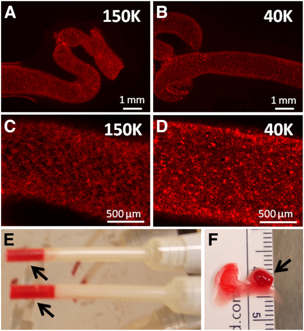
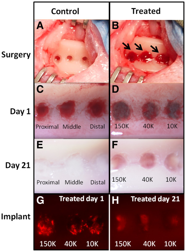
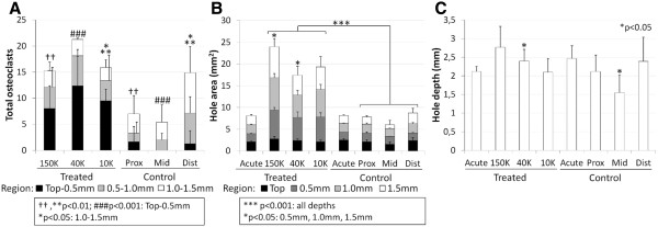
Three microdrill hole defects, 1.4 mm diameter and 2 mm deep, were created in both knee trochlea of 30 month-old New Zealand White rabbits. Each of 3 isotonic chitosan solutions (150, 40, 10 kDa, 80% degree of deaceylation, with fluorescent chitosan tracer) was mixed with autologous rabbit whole blood, clotted with tissue factor to form cylindrical implants, and press-fit in drill holes in the left knee while contralateral holes received tissue factor or no treatment. At day 1 or day 21 post-operative, defects were analyzed by micro-computed tomography, histomorphometry and stereology for bone and soft tissue repair.

RESULTS

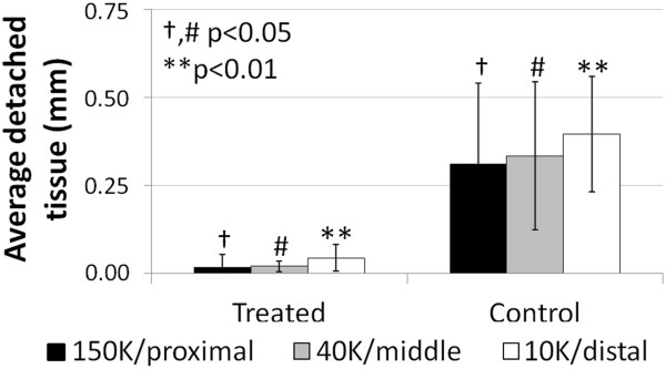
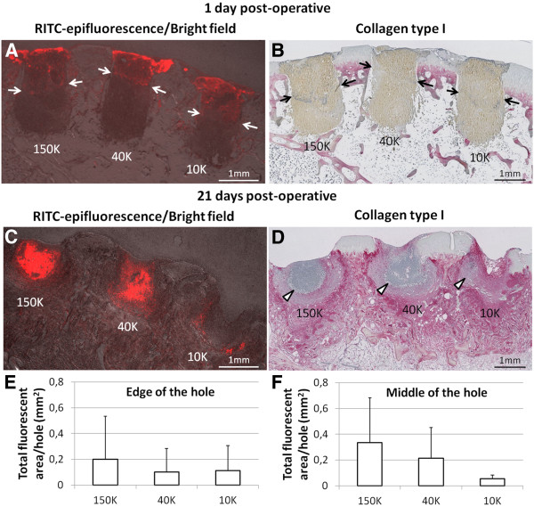
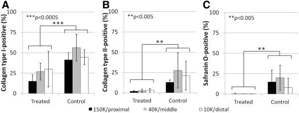
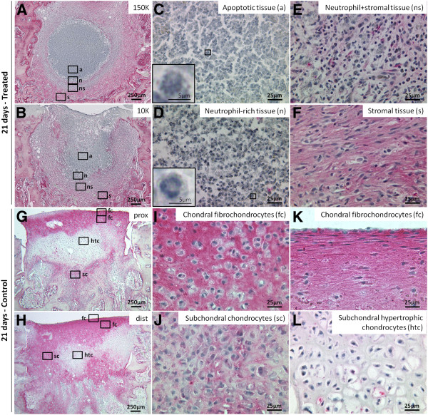
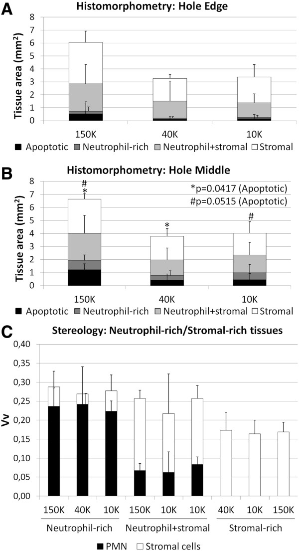
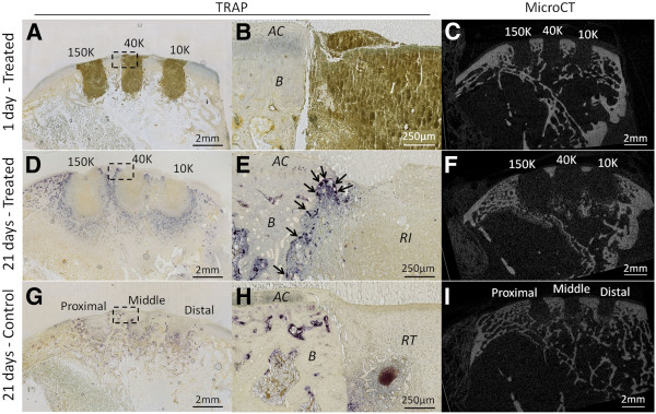
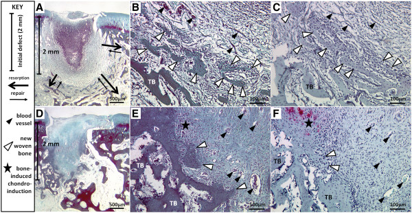
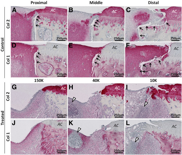
All 3 implants filled the top of defects at day 1 and were partly degraded in situ at 21 days post-operative. All implants attracted neutrophils, osteoclasts and abundant bone marrow-derived stromal cells, stimulated bone resorption followed by new woven bone repair (bone remodeling) and promoted repair tissue-bone integration. 150 kDa chitosan implant was less degraded, and elicited more apoptotic neutrophils and bone resorption than 10 kDa chitosan implant. Drilled controls elicited a poorly integrated fibrous or fibrocartilaginous tissue.

CONCLUSIONS

Pre-solidified implants elicit stromal cells and vigorous bone plate remodeling through a phase involving neutrophil chemotaxis. Pre-solidified chitosan implants are tunable by molecular mass, and could be beneficial for augmented marrow stimulation therapy if the recruited stromal cells can progress to bone and cartilage repair.

摘要

背景

在这项研究中,我们评估了一种新方法,以指导骨髓驱动的关节软骨修复反应在骨骼老化的兔子中。我们假设,植入靠近骨髓的分散壳聚糖颗粒会以分子质量依赖性的方式原位降解,并吸引更多的基质细胞到老年兔子的部位,而不是未治疗对照中的血凝块。

方法

在 30 月龄的新西兰白兔的双侧膝关节滑车中创建了 3 个 1.4 毫米直径和 2 毫米深的微钻孔缺损。将 3 种等渗壳聚糖溶液(150、40、10 kDa,80%去乙酰化度,带有荧光壳聚糖示踪剂)与自体兔全血混合,用组织因子凝结形成圆柱形植入物,并在左侧膝关节的钻孔中压配合,而对侧孔接受组织因子或不治疗。在术后第 1 天或第 21 天,通过微计算机断层扫描、组织形态计量学和体视学分析对骨和软组织修复进行分析。

结果

所有 3 种植入物在第 1 天填充了缺损的顶部,并且在术后第 21 天在原位部分降解。所有植入物都吸引了中性粒细胞、破骨细胞和丰富的骨髓来源的基质细胞,刺激了骨吸收,随后是新的编织骨修复(骨重塑),并促进了修复组织与骨的整合。150 kDa 壳聚糖植入物降解较少,并且比 10 kDa 壳聚糖植入物引起更多的凋亡中性粒细胞和骨吸收。钻孔对照引起了较差整合的纤维或纤维软骨组织。

结论

预固化植入物通过涉及中性粒细胞趋化作用的阶段,募集基质细胞并强烈刺激骨板重塑。预固化壳聚糖植入物可通过分子质量进行调节,如果募集的基质细胞能够进展为骨和软骨修复,那么它们可能对增强骨髓刺激疗法有益。

https://cdn.ncbi.nlm.nih.gov/pmc/blobs/2c0a/3602124/2ca41339d12b/1471-2474-14-27-12.jpg
https://cdn.ncbi.nlm.nih.gov/pmc/blobs/2c0a/3602124/bcce3d24debb/1471-2474-14-27-1.jpg
https://cdn.ncbi.nlm.nih.gov/pmc/blobs/2c0a/3602124/e7694f041d02/1471-2474-14-27-2.jpg
https://cdn.ncbi.nlm.nih.gov/pmc/blobs/2c0a/3602124/7e5ff46c415b/1471-2474-14-27-3.jpg
https://cdn.ncbi.nlm.nih.gov/pmc/blobs/2c0a/3602124/22213067a271/1471-2474-14-27-4.jpg
https://cdn.ncbi.nlm.nih.gov/pmc/blobs/2c0a/3602124/219c08c02a74/1471-2474-14-27-5.jpg
https://cdn.ncbi.nlm.nih.gov/pmc/blobs/2c0a/3602124/f497ef7b450c/1471-2474-14-27-6.jpg
https://cdn.ncbi.nlm.nih.gov/pmc/blobs/2c0a/3602124/a2863b3798ed/1471-2474-14-27-7.jpg
https://cdn.ncbi.nlm.nih.gov/pmc/blobs/2c0a/3602124/106013db3800/1471-2474-14-27-8.jpg
https://cdn.ncbi.nlm.nih.gov/pmc/blobs/2c0a/3602124/7ef5f30e55e6/1471-2474-14-27-9.jpg
https://cdn.ncbi.nlm.nih.gov/pmc/blobs/2c0a/3602124/0af9060d58d2/1471-2474-14-27-10.jpg
https://cdn.ncbi.nlm.nih.gov/pmc/blobs/2c0a/3602124/81bfdd04b341/1471-2474-14-27-11.jpg
https://cdn.ncbi.nlm.nih.gov/pmc/blobs/2c0a/3602124/2ca41339d12b/1471-2474-14-27-12.jpg

相似文献

1
Subchondral pre-solidified chitosan/blood implants elicit reproducible early osteochondral wound-repair responses including neutrophil and stromal cell chemotaxis, bone resorption and repair, enhanced repair tissue integration and delayed matrix deposition.骨下预凝固壳聚糖/血植入物可引发可重复的早期骨软骨创面修复反应,包括中性粒细胞和基质细胞趋化作用、骨质吸收和修复、增强修复组织整合以及延迟基质沉积。
BMC Musculoskelet Disord. 2013 Jan 16;14:27. doi: 10.1186/1471-2474-14-27.
2
Subchondral chitosan/blood implant-guided bone plate resorption and woven bone repair is coupled to hyaline cartilage regeneration from microdrill holes in aged rabbit knees.软骨下壳聚糖/血液植入物引导的骨板吸收和编织骨修复与老年兔膝关节微钻孔处的透明软骨再生相关联。
Osteoarthritis Cartilage. 2014 Feb;22(2):323-33. doi: 10.1016/j.joca.2013.12.011. Epub 2013 Dec 19.
3
Chitosan-glycerol phosphate/blood implants elicit hyaline cartilage repair integrated with porous subchondral bone in microdrilled rabbit defects.壳聚糖-甘油磷酸酯/血液植入物可引发透明软骨修复,并与微钻孔兔缺损处的多孔软骨下骨整合。
Osteoarthritis Cartilage. 2007 Jan;15(1):78-89. doi: 10.1016/j.joca.2006.06.015. Epub 2006 Aug 8.
4
Scaffold-guided subchondral bone repair: implication of neutrophils and alternatively activated arginase-1+ macrophages.支架引导的软骨下骨修复:中性粒细胞和替代性激活的精氨酸酶 1+巨噬细胞的作用。
Am J Sports Med. 2010 Sep;38(9):1845-56. doi: 10.1177/0363546510369547. Epub 2010 Jun 3.
5
Stereological analysis of subchondral angiogenesis induced by chitosan and coagulation factors in microdrilled articular cartilage defects.微钻削关节软骨缺损中壳聚糖和凝血因子诱导的软骨下血管生成的体视学分析。
Osteoarthritis Cartilage. 2013 Jun;21(6):849-59. doi: 10.1016/j.joca.2013.03.012. Epub 2013 Mar 21.
6
Chitosan-glycerol phosphate/blood implants increase cell recruitment, transient vascularization and subchondral bone remodeling in drilled cartilage defects.壳聚糖-甘油磷酸酯/血液植入物可增加钻孔软骨缺损处的细胞募集、短暂血管化和软骨下骨重塑。
Osteoarthritis Cartilage. 2007 Mar;15(3):316-27. doi: 10.1016/j.joca.2006.08.007. Epub 2006 Sep 26.
7
Microdrilled cartilage defects treated with thrombin-solidified chitosan/blood implant regenerate a more hyaline, stable, and structurally integrated osteochondral unit compared to drilled controls.经凝血酶固化壳聚糖/血植入物治疗的微钻取软骨缺损与钻孔对照相比,再生出更透明、稳定且结构整合的软骨-骨单位。
Tissue Eng Part A. 2012 Mar;18(5-6):508-19. doi: 10.1089/ten.TEA.2011.0178. Epub 2011 Nov 11.
8
Acute Osteoclast Activity following Subchondral Drilling Is Promoted by Chitosan and Associated with Improved Cartilage Repair Tissue Integration.软骨下钻孔后破骨细胞活性增强,壳聚糖促进其活性,有助于改善软骨修复组织的整合。
Cartilage. 2011 Apr;2(2):173-85. doi: 10.1177/1947603510381096.
9
Chitosan-glycerol phosphate/blood implants improve hyaline cartilage repair in ovine microfracture defects.壳聚糖-甘油磷酸酯/血液植入物可改善绵羊微骨折缺损处的透明软骨修复。
J Bone Joint Surg Am. 2005 Dec;87(12):2671-2686. doi: 10.2106/JBJS.D.02536.
10
Injectable freeze-dried chitosan-platelet-rich-plasma implants improve marrow-stimulated cartilage repair in a chronic-defect rabbit model.可注射冻干壳聚糖-富血小板血浆植入物改善慢性缺损兔模型中的骨髓刺激软骨修复。
J Tissue Eng Regen Med. 2019 Apr;13(4):599-611. doi: 10.1002/term.2814. Epub 2019 Mar 8.

引用本文的文献

1
Chitosan coatings with distinct innate immune bioactivities differentially stimulate angiogenesis, osteogenesis and chondrogenesis in poly-caprolactone scaffolds with controlled interconnecting pore size.具有独特固有免疫生物活性的壳聚糖涂层,在孔径可控且相互连通的聚己内酯支架中,对血管生成、骨生成和软骨生成有不同程度的刺激作用。
Bioact Mater. 2021 Sep 16;10:430-442. doi: 10.1016/j.bioactmat.2021.09.012. eCollection 2022 Apr.
2
Current Trends in the Evaluation of Osteochondral Lesion Treatments: Histology, Histomorphometry, and Biomechanics in Preclinical Models.当前骨软骨损伤治疗评估的趋势:临床前模型中的组织学、组织形态计量学和生物力学。
Biomed Res Int. 2019 Oct 9;2019:4040236. doi: 10.1155/2019/4040236. eCollection 2019.
3

本文引用的文献

1
Bone-Induced Chondroinduction in Sheep Jamshidi Biopsy Defects with and without Treatment by Subchondral Chitosan-Blood Implant: 1-Day, 3-Week, and 3-Month Repair.羊 Jamshidi 活检缺损中骨诱导性软骨诱导:软骨下壳聚糖-血植入物治疗和不治疗的 1 天、3 周和 3 个月修复。
Cartilage. 2013 Apr;4(2):131-43. doi: 10.1177/1947603512463227.
2
Acute Osteoclast Activity following Subchondral Drilling Is Promoted by Chitosan and Associated with Improved Cartilage Repair Tissue Integration.软骨下钻孔后破骨细胞活性增强,壳聚糖促进其活性,有助于改善软骨修复组织的整合。
Cartilage. 2011 Apr;2(2):173-85. doi: 10.1177/1947603510381096.
3
The cartilage-bone interface.
Effect of a Rapidly Degrading Presolidified 10 kDa Chitosan/Blood Implant and Subchondral Marrow Stimulation Surgical Approach on Cartilage Resurfacing in a Sheep Model.
快速降解的预固化10 kDa壳聚糖/血液植入物及软骨下骨髓刺激手术方法对绵羊模型软骨修复的影响
Cartilage. 2017 Oct;8(4):417-431. doi: 10.1177/1947603516676872. Epub 2016 Nov 11.
4
Knee cartilage defect: marrow stimulating techniques.膝关节软骨缺损:骨髓刺激技术
Curr Rev Musculoskelet Med. 2015 Dec;8(4):451-6. doi: 10.1007/s12178-015-9303-x.
5
A Stereological Method for the Quantitative Evaluation of Cartilage Repair Tissue.一种用于定量评估软骨修复组织的体视学法。
Cartilage. 2015 Apr;6(2):123-32. doi: 10.1177/1947603514560655.
6
Chondrogenic regeneration using bone marrow clots and a porous polycaprolactone-hydroxyapatite scaffold by three-dimensional printing.使用骨髓凝块和通过三维打印制备的多孔聚己内酯-羟基磷灰石支架进行软骨再生。
Tissue Eng Part A. 2015 Apr;21(7-8):1388-97. doi: 10.1089/ten.TEA.2014.0280.
7
Bone substitutes and implantation depths for subchondral bone repair in osteochondral defects of porcine knee joints.猪膝关节骨软骨缺损的软骨下骨修复中骨替代物及植入深度
Knee Surg Sports Traumatol Arthrosc. 2015 May;23(5):1401-1409. doi: 10.1007/s00167-014-2853-4. Epub 2014 Feb 1.
8
Chitosan rate of uptake in HEK293 cells is influenced by soluble versus microparticle state and enhanced by serum-induced cell metabolism and lactate-based media acidification.壳聚糖在 HEK293 细胞中的摄取率受其溶解状态与微颗粒状态的影响,并受血清诱导的细胞代谢和基于乳酸的培养基酸化作用增强。
Molecules. 2013 Jan 15;18(1):1015-35. doi: 10.3390/molecules18011015.
软骨-骨界面
J Knee Surg. 2012 May;25(2):85-97. doi: 10.1055/s-0032-1319782.
4
Microdrilled cartilage defects treated with thrombin-solidified chitosan/blood implant regenerate a more hyaline, stable, and structurally integrated osteochondral unit compared to drilled controls.经凝血酶固化壳聚糖/血植入物治疗的微钻取软骨缺损与钻孔对照相比,再生出更透明、稳定且结构整合的软骨-骨单位。
Tissue Eng Part A. 2012 Mar;18(5-6):508-19. doi: 10.1089/ten.TEA.2011.0178. Epub 2011 Nov 11.
5
Localization of vascular endothelial growth factor during the early stages of osteochondral regeneration using a bioabsorbable synthetic polymer scaffold.在使用可生物吸收的合成聚合物支架进行骨软骨再生的早期阶段,血管内皮生长因子的定位。
J Orthop Res. 2012 Feb;30(2):252-9. doi: 10.1002/jor.21502. Epub 2011 Aug 1.
6
Depth of subchondral perforation influences the outcome of bone marrow stimulation cartilage repair.骨下骨穿孔深度影响骨髓刺激软骨修复的效果。
J Orthop Res. 2011 Aug;29(8):1178-84. doi: 10.1002/jor.21386. Epub 2011 Feb 24.
7
IL-8 and MCP-1 induced by excessive orthodontic force mediates odontoclastogenesis in periodontal tissues.过度正畸力诱导的 IL-8 和 MCP-1 介导牙周组织破骨细胞分化。
Oral Dis. 2011 Jul;17(5):489-98. doi: 10.1111/j.1601-0825.2010.01780.x. Epub 2010 Dec 23.
8
Osteocyte apoptosis is mechanically regulated and induces angiogenesis in vitro.破骨细胞凋亡受机械调控,并在体外诱导血管生成。
J Orthop Res. 2011 Apr;29(4):523-30. doi: 10.1002/jor.21283. Epub 2010 Oct 26.
9
Standardized three-dimensional volumes of interest with adapted surfaces for more precise subchondral bone analyses by micro-computed tomography.标准化的三维感兴趣区与适应表面,用于通过微计算机断层扫描进行更精确的软骨下骨分析。
Tissue Eng Part C Methods. 2011 Apr;17(4):475-84. doi: 10.1089/ten.TEC.2010.0417. Epub 2011 Jan 18.
10
Temporal and spatial modulation of chondrogenic foci in subchondral microdrill holes by chitosan-glycerol phosphate/blood implants.壳聚糖-甘油磷酸/血植入物对软骨下微钻孔中成软骨焦点的时空调节。
Osteoarthritis Cartilage. 2011 Jan;19(1):136-44. doi: 10.1016/j.joca.2010.10.026. Epub 2010 Oct 31.