New Anneliese B, Robin Donald A, Parkinson Amy L, Duffy Joseph R, McNeil Malcom R, Piguet Olivier, Hornberger Michael, Price Cathy J, Eickhoff Simon B, Ballard Kirrie J
Research Imaging Institute, University of Texas Health Science Center San Antonio, San Antonio, TX, USA.
Research Imaging Institute, University of Texas Health Science Center San Antonio, San Antonio, TX, USA ; Department of Neurology, University of Texas Health Science Center San Antonio, San Antonio, TX, USA ; Joint Program in Biomedical Engineering, University of Texas Health Science Center at San Antonio, San Antonio, TX, USA ; Honors College, University of Texas, San Antonio, TX, USA.
Neuroimage Clin. 2015 Mar 25;8:429-39. doi: 10.1016/j.nicl.2015.03.013. eCollection 2015.
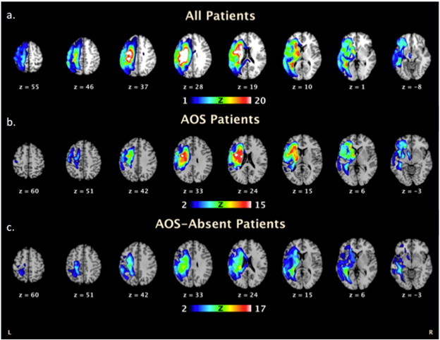
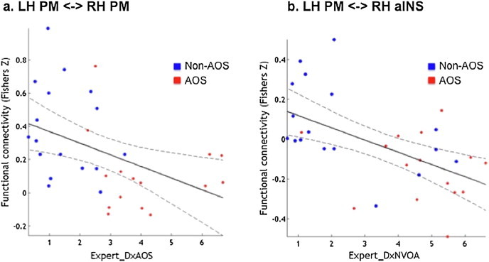
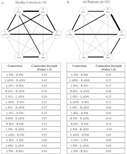
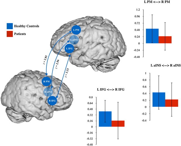
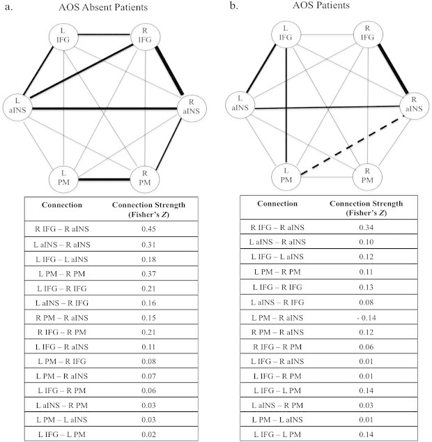
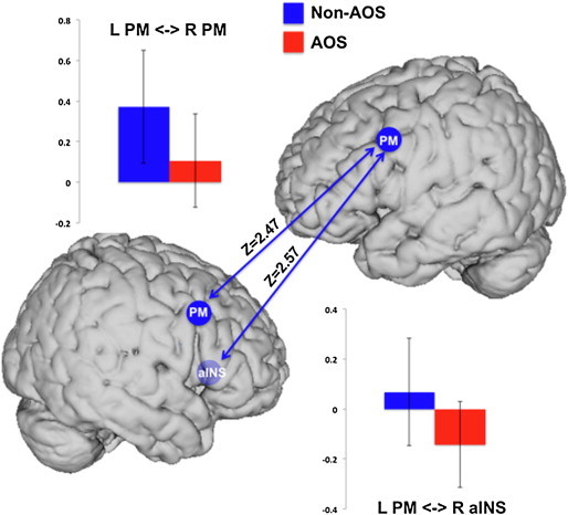
Motor speech disorders, including apraxia of speech (AOS), account for over 50% of the communication disorders following stroke. Given its prevalence and impact, and the need to understand its neural mechanisms, we used resting state functional MRI to examine functional connectivity within a network of regions previously hypothesized as being associated with AOS (bilateral anterior insula (aINS), inferior frontal gyrus (IFG), and ventral premotor cortex (PM)) in a group of 32 left hemisphere stroke patients and 18 healthy, age-matched controls. Two expert clinicians rated severity of AOS, dysarthria and nonverbal oral apraxia of the patients. Fifteen individuals were categorized as AOS and 17 were AOS-absent. Comparison of connectivity in patients with and without AOS demonstrated that AOS patients had reduced connectivity between bilateral PM, and this reduction correlated with the severity of AOS impairment. In addition, AOS patients had negative connectivity between the left PM and right aINS and this effect decreased with increasing severity of non-verbal oral apraxia. These results highlight left PM involvement in AOS, begin to differentiate its neural mechanisms from those of other motor impairments following stroke, and help inform us of the neural mechanisms driving differences in speech motor planning and programming impairment following stroke.
运动性言语障碍,包括言语失用症(AOS),占中风后交流障碍的50%以上。鉴于其患病率和影响,以及了解其神经机制的必要性,我们使用静息态功能磁共振成像来检查一组32名左半球中风患者和18名年龄匹配的健康对照者中,先前假设与AOS相关的区域网络(双侧前岛叶(aINS)、额下回(IFG)和腹侧运动前皮层(PM))内的功能连接。两名专业临床医生对患者的AOS、构音障碍和非言语性口失用症的严重程度进行了评分。15人被归类为AOS患者,17人无AOS。对有和无AOS患者的连接性比较表明,AOS患者双侧PM之间的连接性降低,且这种降低与AOS损伤的严重程度相关。此外,AOS患者左PM与右aINS之间存在负连接,且这种效应随着非言语性口失用症严重程度的增加而降低。这些结果突出了左PM在AOS中的作用,开始将其神经机制与中风后其他运动障碍的神经机制区分开来,并有助于我们了解中风后言语运动计划和编程损伤差异背后的神经机制。