• 文献检索
  • 文档翻译
  • 深度研究
  • 学术资讯
  • Suppr Zotero 插件Zotero 插件
  • 邀请有礼
  • 套餐&价格
  • 历史记录
应用&插件
Suppr Zotero 插件Zotero 插件浏览器插件Mac 客户端Windows 客户端微信小程序
定价
高级版会员购买积分包购买API积分包
服务
文献检索文档翻译深度研究API 文档MCP 服务
关于我们
关于 Suppr公司介绍联系我们用户协议隐私条款
关注我们

Suppr 超能文献

核心技术专利:CN118964589B侵权必究
粤ICP备2023148730 号-1Suppr @ 2026

文献检索

告别复杂PubMed语法,用中文像聊天一样搜索,搜遍4000万医学文献。AI智能推荐,让科研检索更轻松。

立即免费搜索

文件翻译

保留排版,准确专业,支持PDF/Word/PPT等文件格式,支持 12+语言互译。

免费翻译文档

深度研究

AI帮你快速写综述,25分钟生成高质量综述,智能提取关键信息,辅助科研写作。

立即免费体验

图论实际上告诉了我们关于静息态发作间期脑磁图癫痫活动的哪些信息。

What graph theory actually tells us about resting state interictal MEG epileptic activity.

作者信息

Niso Guiomar, Carrasco Sira, Gudín María, Maestú Fernando, Del-Pozo Francisco, Pereda Ernesto

机构信息

Center for Biomedical Technology, Technical University of Madrid, Madrid, Spain ; McConnell Brain Imaging Center, Montreal Neurological Institute, McGill University, Montreal, Canada.

Teaching General Hospital of Ciudad Real, Ciudad Real, Spain.

出版信息

Neuroimage Clin. 2015 May 23;8:503-15. doi: 10.1016/j.nicl.2015.05.008. eCollection 2015.

DOI:10.1016/j.nicl.2015.05.008
PMID:26106575
原文链接:https://pmc.ncbi.nlm.nih.gov/articles/PMC4475779/
Abstract

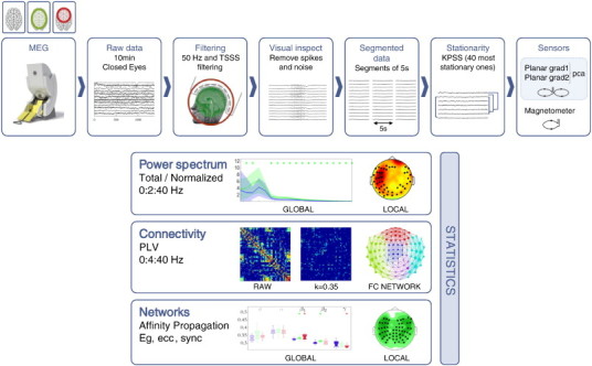
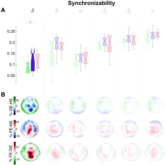
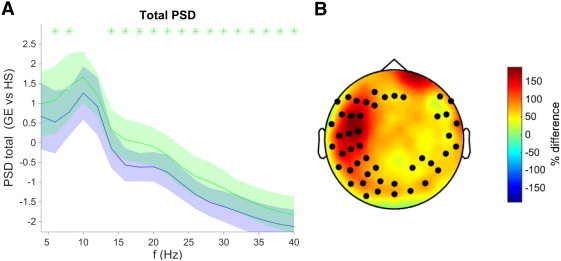
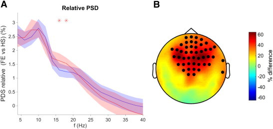
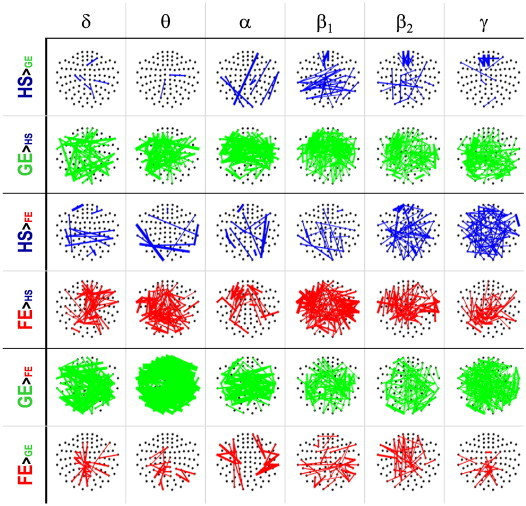
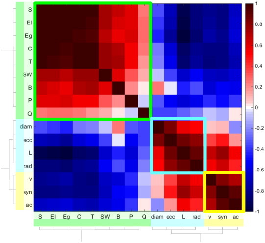
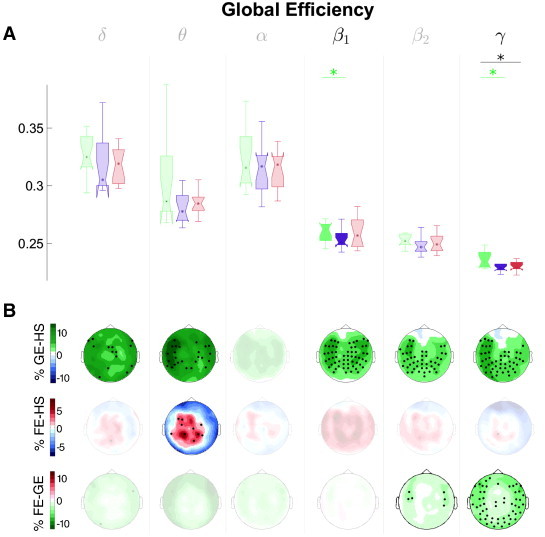
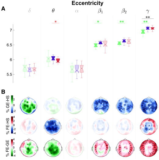
Graph theory provides a useful framework to study functional brain networks from neuroimaging data. In epilepsy research, recent findings suggest that it offers unique insight into the fingerprints of this pathology on brain dynamics. Most studies hitherto have focused on seizure activity during focal epilepsy, but less is known about functional epileptic brain networks during interictal activity in frontal focal and generalized epilepsy. Besides, it is not clear yet which measures are most suitable to characterize these networks. To address these issues, we recorded magnetoencephalographic (MEG) data using two orthogonal planar gradiometers from 45 subjects from three groups (15 healthy controls (7 males, 24 ± 6 years), 15 frontal focal (8 male, 32 ± 16 years) and 15 generalized epileptic (6 male, 27 ± 7 years) patients) during interictal resting state with closed eyes. Then, we estimated the total and relative spectral power of the largest principal component of the gradiometers, and the degree of phase synchronization between each sensor site in the frequency range [0.5-40 Hz]. We further calculated a comprehensive battery of 15 graph-theoretic measures and used the affinity propagation clustering algorithm to elucidate the minimum set of them that fully describe these functional brain networks. The results show that differences in spectral power between the control and the other two groups have a distinctive pattern: generalized epilepsy presents higher total power for all frequencies except the alpha band over a widespread set of sensors; frontal focal epilepsy shows higher relative power in the beta band bilaterally in the fronto-central sensors. Moreover, all network indices can be clustered into three groups, whose exemplars are the global network efficiency, the eccentricity and the synchronizability. Again, the patterns of differences were clear: the brain network of the generalized epilepsy patients presented greater efficiency and lower eccentricity than the control subjects for the high frequency bands, without a clear topography. Besides, the frontal focal epileptic patients showed only reduced eccentricity for the theta band over fronto-temporal and central sensors. These outcomes indicate that functional epileptic brain networks are different to those of healthy subjects during interictal stage at rest, with a unique pattern of dissimilarities for each type of epilepsy. Further, when properly selected, three network indices suffice to provide a comprehensive description of these differences. Yet, since such uniqueness in the pattern of differences is also evident in the power spectrum, we conclude that the added value of the graph theory approach in this context should not be overestimated.

摘要

图论为从神经影像数据研究功能性脑网络提供了一个有用的框架。在癫痫研究中,最近的研究结果表明,它能为这种病理状态在脑动力学上的特征提供独特的见解。迄今为止,大多数研究都集中在局灶性癫痫发作期间的癫痫活动,但对于额叶局灶性癫痫和全身性癫痫发作间期的功能性癫痫脑网络了解较少。此外,目前尚不清楚哪些测量方法最适合表征这些网络。为了解决这些问题,我们使用两个正交平面梯度仪,记录了来自三组45名受试者(15名健康对照者(7名男性,24±6岁)、15名额叶局灶性癫痫患者(8名男性,32±16岁)和15名全身性癫痫患者(6名男性,27±7岁))闭眼休息时的发作间期脑磁图(MEG)数据。然后,我们估计了梯度仪最大主成分的总谱功率和相对谱功率,以及频率范围[0.5 - 40Hz]内每个传感器位点之间的相位同步程度。我们进一步计算了一组15种全面的图论测量指标,并使用亲和传播聚类算法来阐明能完全描述这些功能性脑网络的最小指标集。结果表明,对照组与其他两组之间的谱功率差异具有独特的模式:全身性癫痫在广泛的传感器组中,除α波段外,所有频率的总功率都更高;额叶局灶性癫痫在额中央传感器双侧的β波段显示出更高的相对功率。此外,所有网络指标可分为三组,其典型代表分别是全局网络效率、偏心率和同步性。差异模式再次清晰可见:全身性癫痫患者的脑网络在高频段表现出比对照受试者更高的效率和更低的偏心率,且无明显的拓扑结构。此外,额叶局灶性癫痫患者仅在额颞和中央传感器的θ波段显示出偏心率降低。这些结果表明,在发作间期休息时,功能性癫痫脑网络与健康受试者的不同,每种癫痫类型都有独特的差异模式。此外,当适当选择时,三个网络指标足以全面描述这些差异。然而,由于差异模式中的这种独特性在功率谱中也很明显,我们得出结论,在这种情况下图论方法的附加值不应被高估。

https://cdn.ncbi.nlm.nih.gov/pmc/blobs/2cfe/4475779/a11f27947b67/gr8.jpg
https://cdn.ncbi.nlm.nih.gov/pmc/blobs/2cfe/4475779/45ad1d64ca5e/gr1.jpg
https://cdn.ncbi.nlm.nih.gov/pmc/blobs/2cfe/4475779/3d98d218b7e6/gr2.jpg
https://cdn.ncbi.nlm.nih.gov/pmc/blobs/2cfe/4475779/a9257d070504/gr3.jpg
https://cdn.ncbi.nlm.nih.gov/pmc/blobs/2cfe/4475779/796ca7552734/gr4.jpg
https://cdn.ncbi.nlm.nih.gov/pmc/blobs/2cfe/4475779/c791daf66df5/gr5.jpg
https://cdn.ncbi.nlm.nih.gov/pmc/blobs/2cfe/4475779/147d4ee2f34f/gr6.jpg
https://cdn.ncbi.nlm.nih.gov/pmc/blobs/2cfe/4475779/399415388585/gr7.jpg
https://cdn.ncbi.nlm.nih.gov/pmc/blobs/2cfe/4475779/a11f27947b67/gr8.jpg

相似文献

1
What graph theory actually tells us about resting state interictal MEG epileptic activity.图论实际上告诉了我们关于静息态发作间期脑磁图癫痫活动的哪些信息。
Neuroimage Clin. 2015 May 23;8:503-15. doi: 10.1016/j.nicl.2015.05.008. eCollection 2015.
2
Increased Functional MEG Connectivity as a Hallmark of MRI-Negative Focal and Generalized Epilepsy.功能磁共振脑电连接性增加作为MRI阴性局灶性和全身性癫痫的一个标志
Brain Topogr. 2018 Sep;31(5):863-874. doi: 10.1007/s10548-018-0649-4. Epub 2018 May 15.
3
Abnormal functional brain network in epilepsy patients with focal cortical dysplasia.局灶性皮质发育不良癫痫患者的异常功能性脑网络。
Epilepsy Res. 2014 Nov;108(9):1618-26. doi: 10.1016/j.eplepsyres.2014.09.006. Epub 2014 Sep 16.
4
Altered resting state brain dynamics in temporal lobe epilepsy can be observed in spectral power, functional connectivity and graph theory metrics.颞叶癫痫患者的静息态脑动力学改变可以在频谱功率、功能连接和图论指标中观察到。
PLoS One. 2013 Jul 26;8(7):e68609. doi: 10.1371/journal.pone.0068609. Print 2013.
5
Cortical connectivity in fronto-temporal focal epilepsy from EEG analysis: A study via graph theory.基于图论的脑电分析研究:额颞部局灶性癫痫的皮质连接
Clin Neurophysiol. 2015 Jun;126(6):1108-1116. doi: 10.1016/j.clinph.2014.09.019. Epub 2014 Oct 2.
6
Multi-frequency analysis of brain connectivity networks in migraineurs: a magnetoencephalography study.偏头痛患者脑连接网络的多频分析:一项脑磁图研究
J Headache Pain. 2016;17:38. doi: 10.1186/s10194-016-0636-7. Epub 2016 Apr 18.
7
Non-invasive brain mapping in epilepsy: Applications from magnetoencephalography.癫痫中的无创脑图谱:脑磁图的应用
J Neurosci Methods. 2016 Feb 15;260:283-91. doi: 10.1016/j.jneumeth.2015.11.012. Epub 2015 Nov 28.
8
Lateralization of epilepsy using intra-hemispheric brain networks based on resting-state MEG data.基于静息态脑磁图数据的半球内脑网络对癫痫的侧化研究。
Hum Brain Mapp. 2020 Aug 1;41(11):2964-2979. doi: 10.1002/hbm.24990. Epub 2020 May 13.
9
State dependent properties of epileptic brain networks: comparative graph-theoretical analyses of simultaneously recorded EEG and MEG.癫痫脑网络的状态依赖性特性:同时记录的 EEG 和 MEG 的比较图论分析。
Clin Neurophysiol. 2010 Feb;121(2):172-85. doi: 10.1016/j.clinph.2009.10.013. Epub 2009 Dec 31.
10
Increased segregation of brain networks in focal epilepsy: An fMRI graph theory finding.局灶性癫痫中脑网络分离增加:一项功能磁共振成像图论研究发现。
Neuroimage Clin. 2015 May 22;8:536-42. doi: 10.1016/j.nicl.2015.05.009. eCollection 2015.

引用本文的文献

1
Quantitative analysis of the resting-state EEG power spectrum in patients with epilepsy comorbid with anxiety and depression.癫痫合并焦虑和抑郁患者静息态脑电图功率谱的定量分析。
Acta Epileptol. 2025 Mar 31;7(1):22. doi: 10.1186/s42494-025-00206-6.
2
Frequency-band specific directed connectivity networks reveal functional disruptions and pathogenic patterns in temporal lobe epilepsy: a MEG study.特定频段定向连接网络揭示颞叶癫痫的功能破坏和致病模式:一项脑磁图研究
Sci Rep. 2025 Apr 10;15(1):12326. doi: 10.1038/s41598-025-90299-3.
3
Symptom network analysis of prefrontal seizures.

本文引用的文献

1
OMEGA: The Open MEG Archive.欧米伽:开放式脑磁图存档库。
Neuroimage. 2016 Jan 1;124(Pt B):1182-1187. doi: 10.1016/j.neuroimage.2015.04.028. Epub 2015 Apr 18.
2
Individual diversity of functional brain network economy.功能性脑网络经济性的个体差异。
Brain Connect. 2015 Apr;5(3):156-65. doi: 10.1089/brain.2014.0306. Epub 2014 Dec 29.
3
Network dynamics of the brain and influence of the epileptic seizure onset zone.大脑的网络动力学与癫痫发作起始区的影响。
前额叶癫痫的症状网络分析
Epilepsia. 2025 Jul;66(7):2566-2577. doi: 10.1111/epi.18372. Epub 2025 Mar 19.
4
Altered Directed-Connectivity Network in Temporal Lobe Epilepsy: A MEG Study.颞叶癫痫中定向连接网络的改变:一项脑磁图研究
Sensors (Basel). 2025 Feb 22;25(5):1356. doi: 10.3390/s25051356.
5
Quantitative EEG signatures in patients with and without epilepsy development after a first seizure.首次发作后有或无癫痫发展患者的定量脑电图特征。
Epilepsia Open. 2025 Apr;10(2):427-440. doi: 10.1002/epi4.13128. Epub 2025 Mar 4.
6
Structural connectome constrained graphical lasso for MEG partial coherence.用于脑磁图部分相干性的结构连接组约束图模型选择法
Netw Neurosci. 2022 Oct 1;6(4):1219-1242. doi: 10.1162/netn_a_00267. eCollection 2022.
7
Exploring brain network oscillations during seizures in drug-naïve patients with juvenile absence epilepsy.探索初治青少年失神癫痫患者癫痫发作期间的脑网络振荡。
Front Neurol. 2024 Mar 14;15:1340959. doi: 10.3389/fneur.2024.1340959. eCollection 2024.
8
Epilepsy-related functional brain network alterations are already present at an early age in the GAERS rat model of genetic absence epilepsy.在遗传性失神癫痫的GAERS大鼠模型中,与癫痫相关的功能性脑网络改变在幼年时就已存在。
Front Neurol. 2024 Mar 11;15:1355862. doi: 10.3389/fneur.2024.1355862. eCollection 2024.
9
Disentanglement of Resting State Brain Networks for Localizing Epileptogenic Zone in Focal Epilepsy.分离静息态脑网络以定位局灶性癫痫的致痫区。
Brain Topogr. 2024 Jan;37(1):152-168. doi: 10.1007/s10548-023-01025-z. Epub 2023 Dec 19.
10
An individual data-driven virtual resection model based on epileptic network dynamics in children with intractable epilepsy: a magnetoencephalography interictal activity application.基于难治性癫痫患儿癫痫网络动力学的个体数据驱动虚拟切除模型:脑磁图发作间期活动应用
Brain Commun. 2023 May 25;5(3):fcad168. doi: 10.1093/braincomms/fcad168. eCollection 2023.
Proc Natl Acad Sci U S A. 2014 Dec 9;111(49):E5321-30. doi: 10.1073/pnas.1401752111. Epub 2014 Nov 17.
4
Revealing a brain network endophenotype in families with idiopathic generalised epilepsy.揭示特发性全身性癫痫家族中的脑网络内表型。
PLoS One. 2014 Oct 10;9(10):e110136. doi: 10.1371/journal.pone.0110136. eCollection 2014.
5
Can spurious indications for phase synchronization due to superimposed signals be avoided?由叠加信号导致的相位同步的虚假指示能否避免?
Chaos. 2014 Sep;24(3):033112. doi: 10.1063/1.4890568.
6
Modern network science of neurological disorders.神经系统疾病的现代网络科学。
Nat Rev Neurosci. 2014 Oct;15(10):683-95. doi: 10.1038/nrn3801. Epub 2014 Sep 4.
7
Structural connectivity based whole brain modelling in epilepsy.癫痫中基于结构连接性的全脑建模
J Neurosci Methods. 2014 Oct 30;236:51-7. doi: 10.1016/j.jneumeth.2014.08.010. Epub 2014 Aug 19.
8
Clinical correlates of graph theory findings in temporal lobe epilepsy.颞叶癫痫中图论研究结果的临床相关性。
Seizure. 2014 Nov;23(10):809-18. doi: 10.1016/j.seizure.2014.07.004. Epub 2014 Jul 23.
9
Network Connectivity in Epilepsy: Resting State fMRI and EEG-fMRI Contributions.癫痫中的网络连通性:静息态功能磁共振成像和脑电图-功能磁共振成像的贡献
Front Neurol. 2014 Jul 4;5:93. doi: 10.3389/fneur.2014.00093. eCollection 2014.
10
Frequency domain beamforming of magnetoencephalographic beta band activity in epilepsy patients with focal cortical dysplasia.局灶性皮质发育不良癫痫患者脑磁图β频段活动的频域波束形成
Epilepsy Res. 2014 Sep;108(7):1195-203. doi: 10.1016/j.eplepsyres.2014.05.003. Epub 2014 May 13.