Bénézech Cécile, Luu Nguyet-Thin, Walker Jennifer A, Kruglov Andrei A, Loo Yunhua, Nakamura Kyoko, Zhang Yang, Nayar Saba, Jones Lucy H, Flores-Langarica Adriana, McIntosh Alistair, Marshall Jennifer, Barone Francesca, Besra Gurdyal, Miles Katherine, Allen Judith E, Gray Mohini, Kollias George, Cunningham Adam F, Withers David R, Toellner Kai Michael, Jones Nick D, Veldhoen Marc, Nedospasov Sergei A, McKenzie Andrew N J, Caamaño Jorge H
School of Immunity and Infection, IBR-MRC Centre for Immune Regulation, College of Medical and Dental Sciences, University of Birmingham, Birmingham, B15 2TT, UK.
MRC Laboratory of Molecular Biology, Cambridge, UK.
Nat Immunol. 2015 Aug;16(8):819-828. doi: 10.1038/ni.3215. Epub 2015 Jun 29.
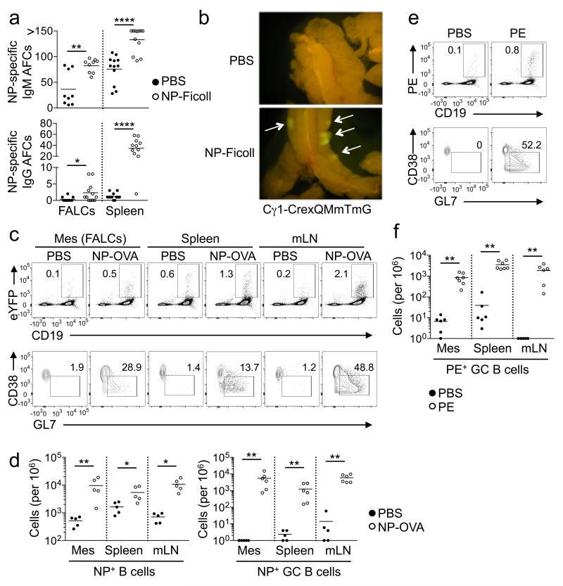
Fat-associated lymphoid clusters (FALCs) are a type of lymphoid tissue associated with visceral fat. Here we found that the distribution of FALCs was heterogeneous, with the pericardium containing large numbers of these clusters. FALCs contributed to the retention of B-1 cells in the peritoneal cavity through high expression of the chemokine CXCL13, and they supported B cell proliferation and germinal center differentiation during peritoneal immunological challenges. FALC formation was induced by inflammation, which triggered the recruitment of myeloid cells that expressed tumor-necrosis factor (TNF) necessary for signaling via the TNF receptors in stromal cells. Natural killer T cells (NKT cells) restricted by the antigen-presenting molecule CD1d were likewise required for the inducible formation of FALCs. Thus, FALCs supported and coordinated the activation of innate B cells and T cells during serosal immune responses.
脂肪相关淋巴簇(FALCs)是一种与内脏脂肪相关的淋巴组织。在此我们发现,FALCs的分布是异质性的,心包中含有大量此类簇。FALCs通过趋化因子CXCL13的高表达促使B-1细胞保留在腹腔中,并且在腹膜免疫应激期间支持B细胞增殖和生发中心分化。炎症诱导FALC形成,炎症触发髓样细胞募集,这些髓样细胞表达基质细胞中通过肿瘤坏死因子(TNF)受体进行信号传导所必需的TNF。受抗原呈递分子CD1d限制的自然杀伤T细胞(NKT细胞)同样是FALCs诱导形成所必需的。因此,FALCs在浆膜免疫反应期间支持并协调固有B细胞和T细胞的激活。