Massey Andrew J
Vernalis Research, Granta Park, Cambridge, CB21 6GB, United Kingdom.
PLoS One. 2015 Jul 28;10(7):e0134306. doi: 10.1371/journal.pone.0134306. eCollection 2015.
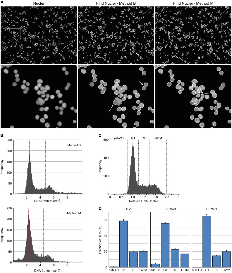
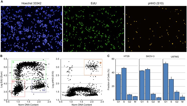
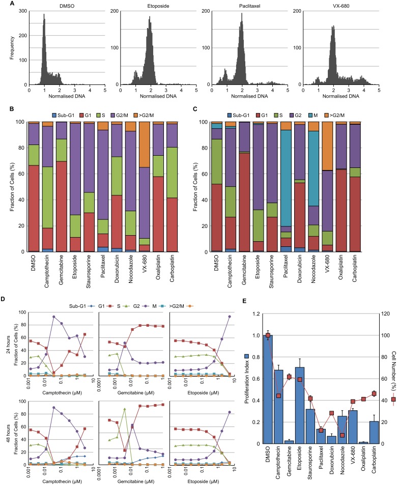
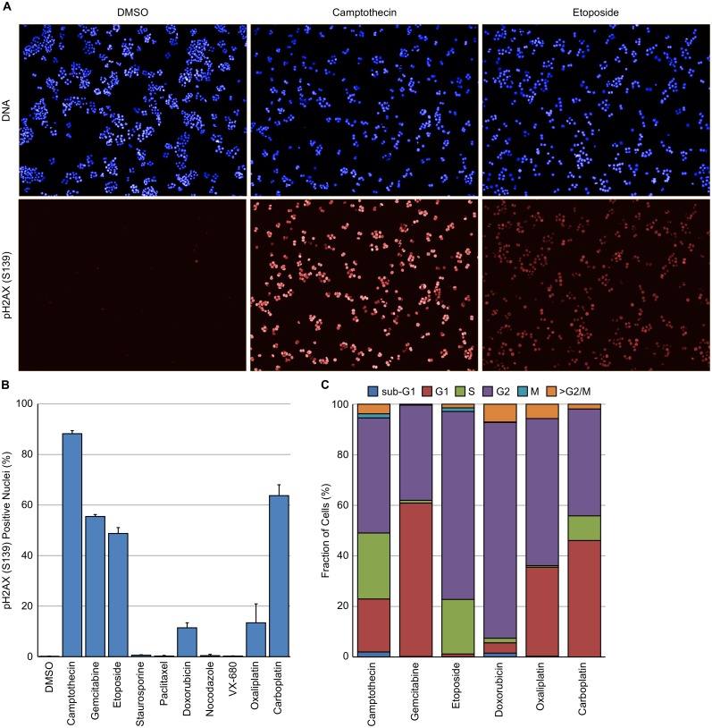
High-content imaging is a powerful tool for determining cell phenotypes at the single cell level. Characterising the effect of small molecules on cell cycle distribution is important for understanding their mechanism of action especially in oncology drug discovery but also for understanding potential toxicology liabilities. Here, a high-throughput phenotypic assay utilising the PerkinElmer Operetta high-content imager and Harmony software to determine cell cycle distribution is described. PhenoLOGIC, a machine learning algorithm within Harmony software was employed to robustly separate single cells from cell clumps. DNA content, EdU incorporation and pHH3 (S10) expression levels were subsequently utilised to separate cells into the various phases of the cell cycle. The assay is amenable to multiplexing with an additional pharmacodynamic marker to assess cell cycle changes within a specific cellular sub-population. Using this approach, the cell cycle distribution of γH2AX positive nuclei was determined following treatment with DNA damaging agents. Likewise, the assay can be multiplexed with Ki67 to determine the fraction of quiescent cells and with BrdU dual labelling to determine S-phase duration. This methodology therefore provides a relatively cheap, quick and high-throughput phenotypic method for determining accurate cell cycle distribution for small molecule mechanism of action and drug toxicity studies.
高内涵成像技术是在单细胞水平确定细胞表型的强大工具。表征小分子对细胞周期分布的影响对于理解其作用机制至关重要,尤其在肿瘤学药物研发中,同时对于了解潜在的毒理学风险也很重要。在此,描述了一种利用珀金埃尔默Operetta高内涵成像仪和Harmony软件来确定细胞周期分布的高通量表型分析方法。Harmony软件中的机器学习算法PhenoLOGIC被用于从细胞团块中稳健地分离单个细胞。随后利用DNA含量、EdU掺入和pHH3(S10)表达水平将细胞分离到细胞周期的各个阶段。该分析方法适合与额外的药效学标记物进行多重分析,以评估特定细胞亚群内的细胞周期变化。采用这种方法,在用DNA损伤剂处理后确定了γH2AX阳性细胞核的细胞周期分布。同样,该分析方法可与Ki67进行多重分析以确定静止细胞的比例,并与BrdU双重标记以确定S期持续时间。因此,该方法为小分子作用机制和药物毒性研究中确定准确的细胞周期分布提供了一种相对廉价、快速且高通量的表型分析方法。