Gali Julio Cesar, Resina André França, Pedro Gabriel, Neto Ildefonso Angelo Mora, Almagro Marco Antonio Pires, da Silva Phelipe Augusto Cintra, Caetano Edie Benedito
Orthopedics and Traumatology Service, School of Medical Sciences and Health of Sorocaba, Pontifical Catholic University of São Paulo (PUC-SP), Sorocaba, SP, Brazil.
Rev Bras Ortop. 2014 Oct 27;49(6):625-9. doi: 10.1016/j.rboe.2013.10.004. eCollection 2014 Nov-Dec.
To describe the path of the infrapatellar branch of the saphenous nerve (IBSN) using the medial joint line, anterior tibial tuberosity (ATT), tibial collateral ligament and a horizontal line parallel to the medial joint line that passes over the ATT, as reference points, in order to help surgeons to diminish the likelihood of injuring this nerve branch during reconstruction of the anterior cruciate ligament (ACL) using flexor tendons.
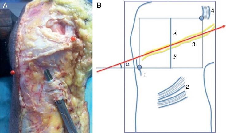
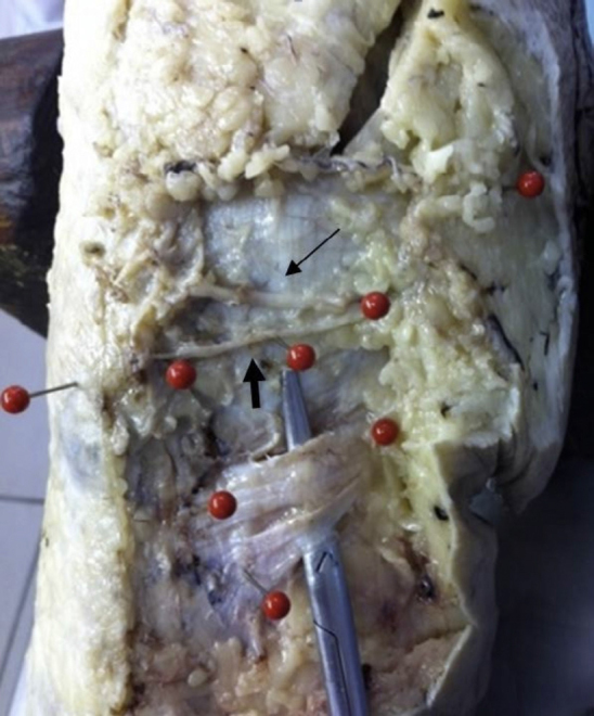
Ten frozen knees that originated from amputations were examined. Through anatomical dissection performed with the specimens flexed, we sought to find the IBSN, from its most medial and proximal portion to its most lateral and distal portion. Following this, the anatomical specimens were photographed and, using the ImageJ software, we determined the distance from the IBSN to the medial joint line and to a lower horizontal line going through the ATT and parallel to the first line. We also measured the angle of the direction of the path of the nerve branch in relation to this lower line.
The mean angle of the path of the nerve branch in relation to the lower horizontal line was 17.50 ± 6.17°. The mean distance from the IBSN to the medial joint line was 2.61 ± 0.59 cm and from the IBSN to the lower horizontal line, 1.44 ± 0.51 cm.
The IBSN was found in all the knees studied. In three knees, we found a second branch proximal to the first one. The direction of its path was always from proximal and medial to distal and lateral. The IBSN was always proximal and medial to the ATT and distal to the medial joint line. The medial angle between its direction and a horizontal line going through the ATT was 17.50 ± 6.17°.
以膝关节内侧关节线、胫骨结节前侧(ATT)、胫侧副韧带以及一条与内侧关节线平行且经过ATT的水平线作为参考点,描述隐神经髌下支(IBSN)的走行路径,以帮助外科医生在使用屈肌腱重建前交叉韧带(ACL)过程中降低损伤该神经分支的可能性。
检查10个因截肢而获取的冷冻膝关节。在标本屈曲状态下进行解剖,从IBSN最内侧和近端部分直至其最外侧和远端部分寻找该神经。之后,对解剖标本进行拍照,并使用ImageJ软件确定IBSN到内侧关节线以及到经过ATT且与第一条线平行的下方水平线的距离。我们还测量了神经分支走行方向与该下方线的夹角。
神经分支走行方向与下方水平线的平均夹角为17.50±6.17°。IBSN到内侧关节线的平均距离为2.61±0.59厘米,到下方水平线的平均距离为1.44±0.51厘米。
在所研究的所有膝关节中均发现了IBSN。在3个膝关节中,我们在第一个分支近端发现了第二个分支。其走行方向始终是从近端和内侧到远端和外侧。IBSN始终位于ATT的近端和内侧以及内侧关节线的远端。其走行方向与经过ATT的水平线之间的内侧夹角为17.50±6.17°。