Adriaenssens Alice, Lam Brian Yee Hong, Billing Lawrence, Skeffington Katie, Sewing Sabine, Reimann Frank, Gribble Fiona
Metabolic Research Laboratories (A.A., B.Y.H.L., L.B., K.S., F.R., F.G.), Wellcome Trust-Medical Research Council Institute of Metabolic Science, Addenbrooke's Hospital, Cambridge CB2 0QQ, United Kingdom; and Pharma Research and Early Development (S.S.), Roche Innovation Center Basel, F. Hoffmann-La Roche AG, 4070 Basel, Switzerland.
Endocrinology. 2015 Nov;156(11):3924-36. doi: 10.1210/en.2015-1301. Epub 2015 Aug 4.
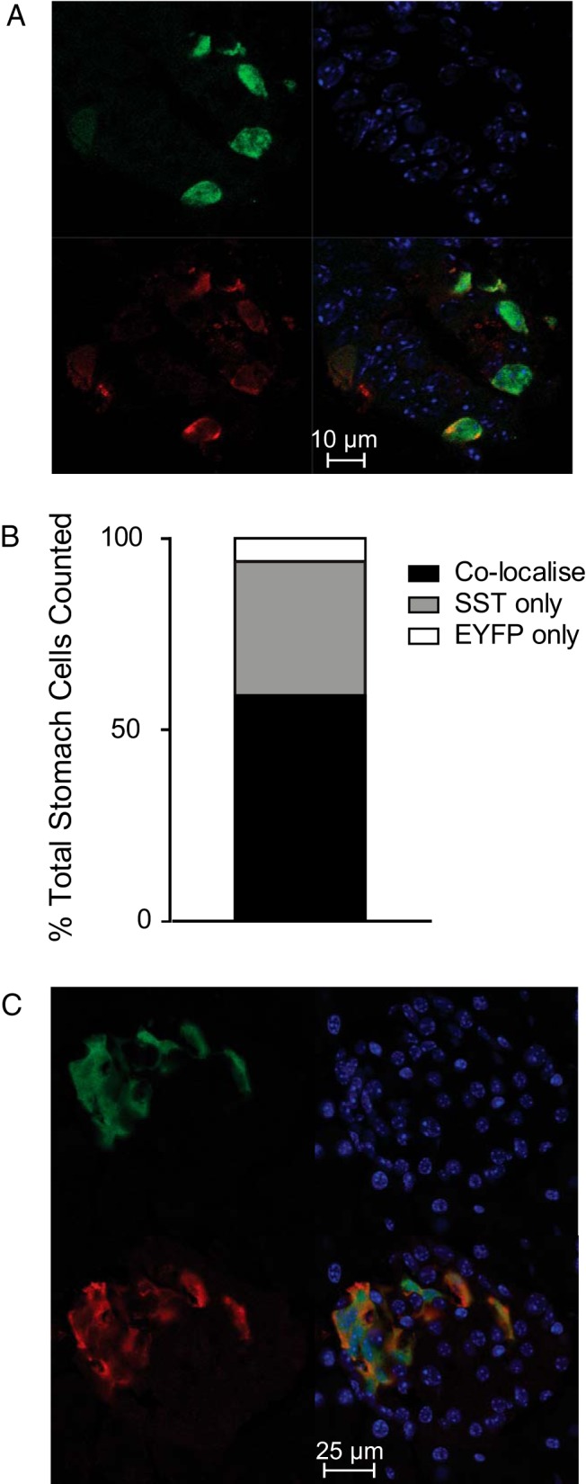
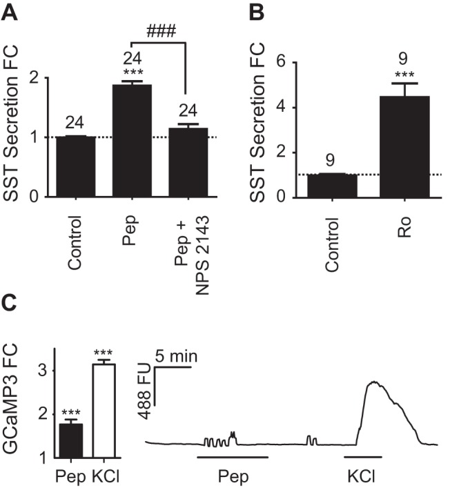
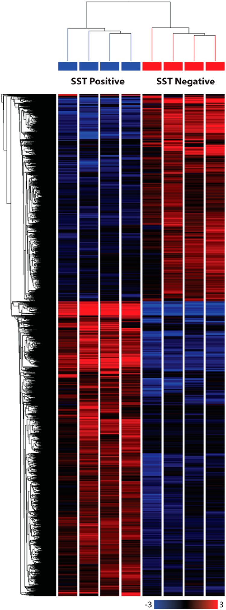
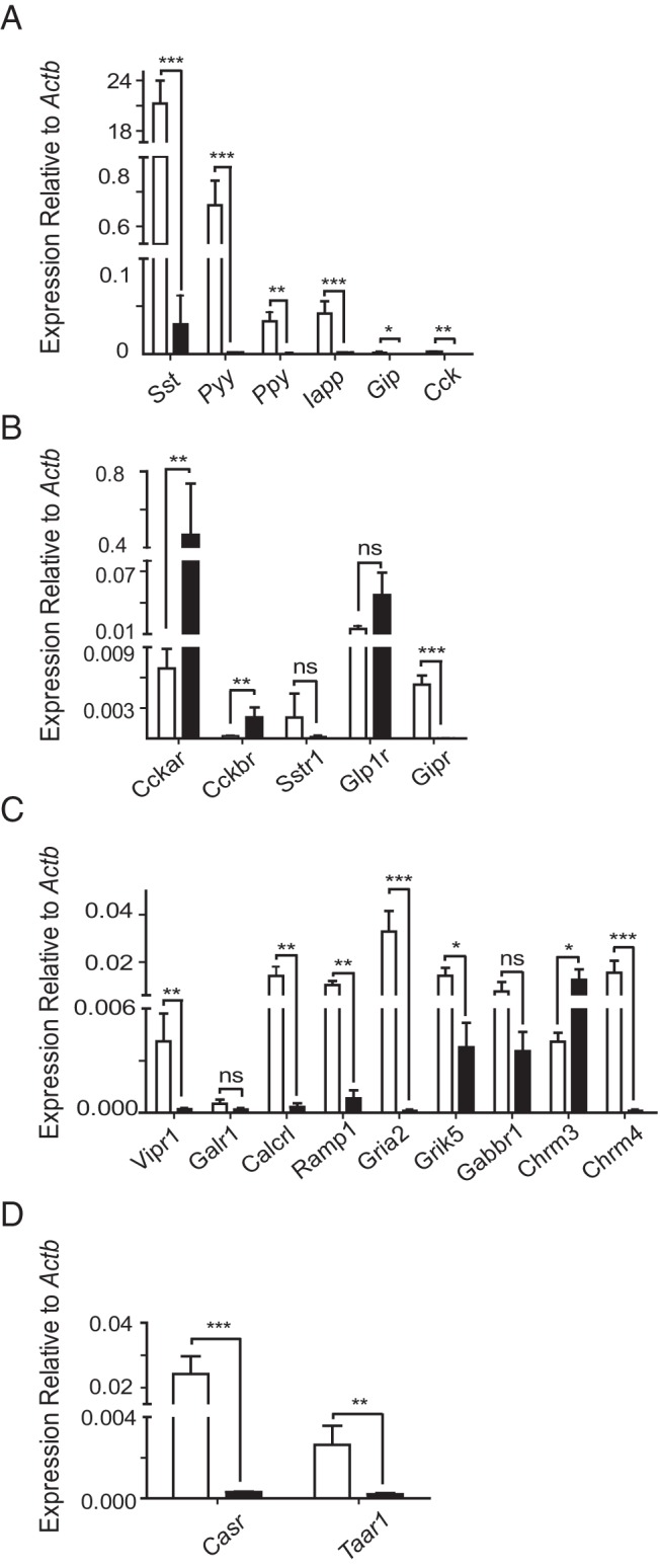
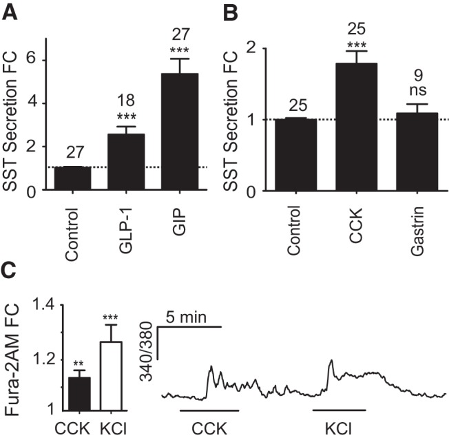
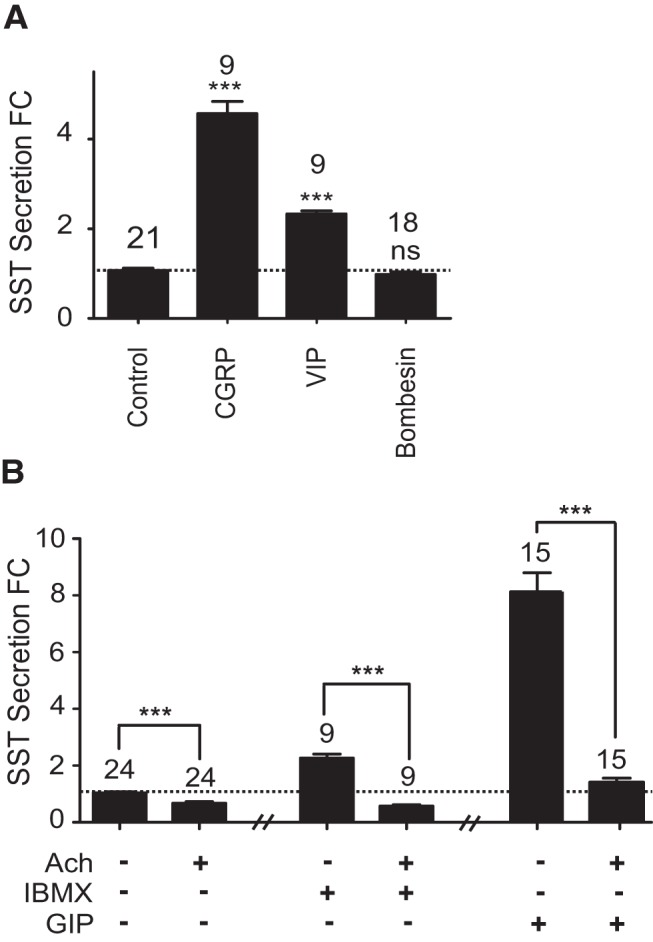
The stomach epithelium contains a myriad of enteroendocrine cells that modulate a range of physiological functions, including postprandial secretion of regulatory peptides, gastric motility, and nutrient absorption. Somatostatin (SST)-producing D-cells are present in the oxyntic and pyloric regions of the stomach, and provide a tonic inhibitory tone that regulates activity of neighboring enteroendocrine cells and gastric acid secretion. Cellular mechanisms underlying the effects of regulatory factors on gastric D-cells are poorly defined due to problems in identifying primary D-cells, and uncertainty remains about which stimuli influence D-cells directly. In this study, we introduce a transgenic mouse line, SST-Cre, which upon crossing with Cre reporter strains, facilitates the identification and purification of gastric D-cells, or cell-specific expression of genetically encoded calcium indicators. Populations of D-cells from the gastric antrum and corpus were isolated and analyzed by RNA sequencing and quantitative RT-PCR. The expression of hormones, hormone receptors, neurotransmitter receptors, and nutrient receptors was quantified. Pyy, Gipr, Chrm4, Calcrl, Taar1, and Casr were identified as genes that are highly enriched in D-cells compared with SST-negative cells. Hormone secretion assays performed in mixed gastric epithelial cultures confirmed that SST secretion is regulated by incretin hormones, cholecystokinin, acetylcholine, vasoactive intestinal polypeptide, calcitonin gene-related polypeptide, oligopetides, and trace amines. Cholecystokinin and oligopeptides elicited increases in intracellular calcium in single-cell imaging experiments performed using cultured D-cells. Our data provide the first transcriptomic analysis and functional characterization of gastric D-cells, and identify regulatory pathways that underlie the direct detection of stimuli by this cell type.
胃上皮含有大量肠内分泌细胞,这些细胞调节一系列生理功能,包括餐后调节肽的分泌、胃动力和营养吸收。产生生长抑素(SST)的D细胞存在于胃的泌酸区和幽门区,并提供一种紧张性抑制作用,调节相邻肠内分泌细胞的活性和胃酸分泌。由于在鉴定原代D细胞方面存在问题,调节因子对胃D细胞作用的细胞机制尚不清楚,并且对于哪些刺激直接影响D细胞仍存在不确定性。在本研究中,我们引入了一种转基因小鼠品系SST-Cre,它与Cre报告菌株杂交后,有助于胃D细胞的鉴定和纯化,或基因编码钙指示剂的细胞特异性表达。通过RNA测序和定量RT-PCR对来自胃窦和胃体的D细胞群体进行分离和分析。对激素、激素受体、神经递质受体和营养受体的表达进行了定量。与SST阴性细胞相比,Pyy、Gipr、Chrm4、Calcrl、Taar1和Casr被鉴定为在D细胞中高度富集的基因。在混合胃上皮培养物中进行的激素分泌试验证实,SST分泌受肠促胰岛素激素、胆囊收缩素、乙酰胆碱、血管活性肠多肽、降钙素基因相关多肽、寡肽和痕量胺的调节。在使用培养的D细胞进行的单细胞成像实验中,胆囊收缩素和寡肽引起细胞内钙增加。我们的数据提供了胃D细胞的首次转录组分析和功能表征,并确定了该细胞类型直接检测刺激的调节途径。