Chung Seyung, Dwabe Sami, Elshimali Yayha, Sukhija Hemlata, Aroh Clement, Vadgama Jaydutt V
Division of Cancer Research and Training, Department of Medicine, Charles R. Drew University of Medicine and Science, 1731 120th street, Los Angeles, California, 90059, United States of America.
Division of Cancer Research and Training, Department of Medicine, Charles R. Drew University of Medicine and Science, 1731 120th street, Los Angeles, California, 90059, United States of America; David Geffen UCLA School of Medicine, University of California Los Angeles, Los Angeles, California, United States of America.
PLoS One. 2015 Aug 10;10(8):e0134948. doi: 10.1371/journal.pone.0134948. eCollection 2015.
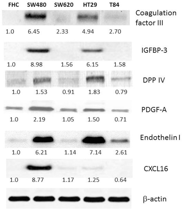
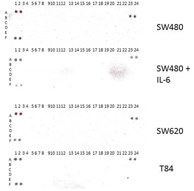
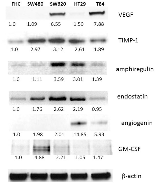
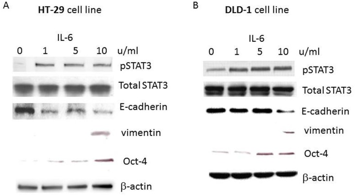
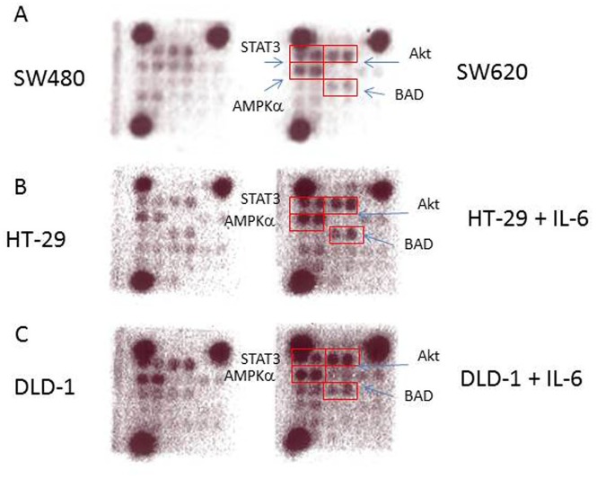
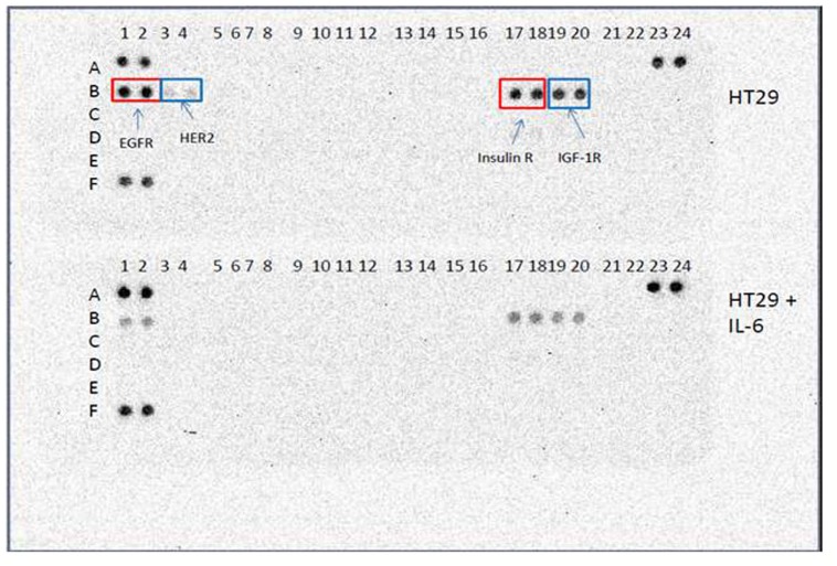
Colorectal cancer (CRC) is one of the three leading causes for cancer mortality. CRC kills over 600,000 people annually worldwide. The most common cause of death from CRC is the metastasis to distant organs. However, biomarkers for CRC metastasis remain ill-defined. We compared primary and metastatic CRC cell lines for their angiogenesis-protein profiles and intracellular signaling profiles to identify novel biomarkers for CRC metastasis. To this end, we used primary and metastatic CRC cell lines as a model system and normal human colon cell line as a control. The angiogenesis profiles two isogenic CRC cell lines, SW480 and SW620, and HT-29 and T84 revealed that VEGF was upregulated in both SW620 and T84 whereas coagulation factor III, IGFBP-3, DPP IV, PDGF AA/AB, endothelin I and CXCL16 were downregulated specifically in metastatic cell lines. Furthermore, we found that TIMP-1, amphiregulin, endostatin, angiogenin were upregulated in SW620 whereas downregulated in T84. Angiogenin was downregulated in T84 and GM-CSF was also downregulated in SW620. To induce CRC cell metastasis, we treated cells with pro-inflammatory cytokine IL-6. Upon IL-6 treatment, epithelial-mesenchymal transition was induced in CRC cells. When DLD-1 and HT-29 cells were treated with IL-6; Akt, STAT3, AMPKα and Bad phosphorylation levels were increased. Interestingly, SW620 showed the same signal activation pattern with IL-6 treatment of HT-29 and DLD-1. Our data suggest that Akt, STAT3, AMPKα and Bad activation can be biomarkers for metastatic colorectal cancer. IL-6 treatment specifically reduced phosphorylation levels of EGFR, HER2 receptor, Insulin R and IGF-1R in receptor tyrosine kinase array study with HT-29. Taken together, we have identified novel biomarkers for metastatic CRC through the angiogenesis-antibody array and intracellular signaling array studies. Present study suggests that those novel biomarkers can be used as CRC prognosis biomarkers, and as potential targets for the metastatic CRC therapy.
结直肠癌(CRC)是癌症死亡的三大主要原因之一。全球每年有超过60万人死于CRC。CRC最常见的死亡原因是远处器官转移。然而,CRC转移的生物标志物仍不明确。我们比较了原发性和转移性CRC细胞系的血管生成蛋白谱和细胞内信号谱,以确定CRC转移的新型生物标志物。为此,我们使用原发性和转移性CRC细胞系作为模型系统,以正常人结肠细胞系作为对照。对两个同基因CRC细胞系SW480和SW620以及HT-29和T84的血管生成谱分析显示,VEGF在SW620和T84中均上调,而凝血因子III、IGFBP-3、DPP IV、PDGF AA/AB、内皮素I和CXCL16在转移性细胞系中特异性下调。此外,我们发现TIMP-1、双调蛋白、内皮抑素、血管生成素在SW620中上调而在T84中下调。血管生成素在T84中下调,GM-CSF在SW620中也下调。为了诱导CRC细胞转移,我们用促炎细胞因子IL-6处理细胞。经IL-6处理后,CRC细胞发生上皮-间质转化。当DLD-1和HT-29细胞用IL-6处理时,Akt、STAT3、AMPKα和Bad的磷酸化水平升高。有趣的是,SW620在接受IL-6处理时与HT-29和DLD-1表现出相同的信号激活模式。我们的数据表明,Akt、STAT3、AMPKα和Bad的激活可能是转移性结直肠癌的生物标志物。在对HT-29进行的受体酪氨酸激酶阵列研究中,IL-6处理特异性降低了EGFR、HER2受体、胰岛素R和IGF-1R的磷酸化水平。综上所述,我们通过血管生成抗体阵列和细胞内信号阵列研究确定了转移性CRC的新型生物标志物。目前的研究表明,这些新型生物标志物可作为CRC预后生物标志物,以及转移性CRC治疗的潜在靶点。