Mikoliunaite Lina, Rodriguez Raul D, Sheremet Evgeniya, Kolchuzhin Vladimir, Mehner Jan, Ramanavicius Arunas, Zahn Dietrich R T
Department of Physical Chemistry, Vilnius University, Vilnius, Lithuania.
Semiconductor Physics, Technische Universität Chemnitz, Chemnitz, Germany.
Sci Rep. 2015 Aug 27;5:13150. doi: 10.1038/srep13150.
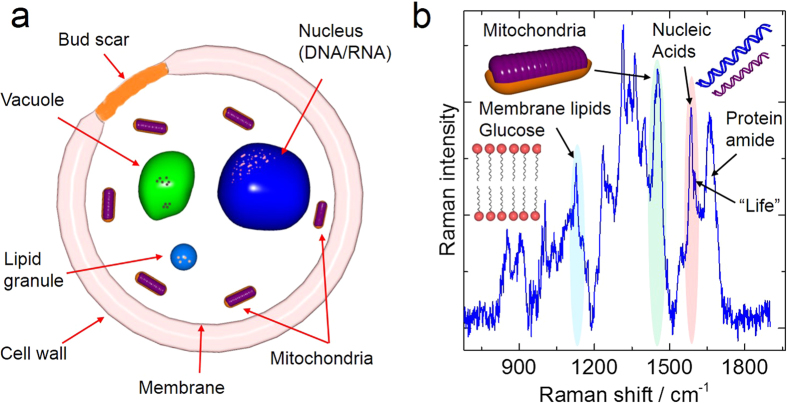
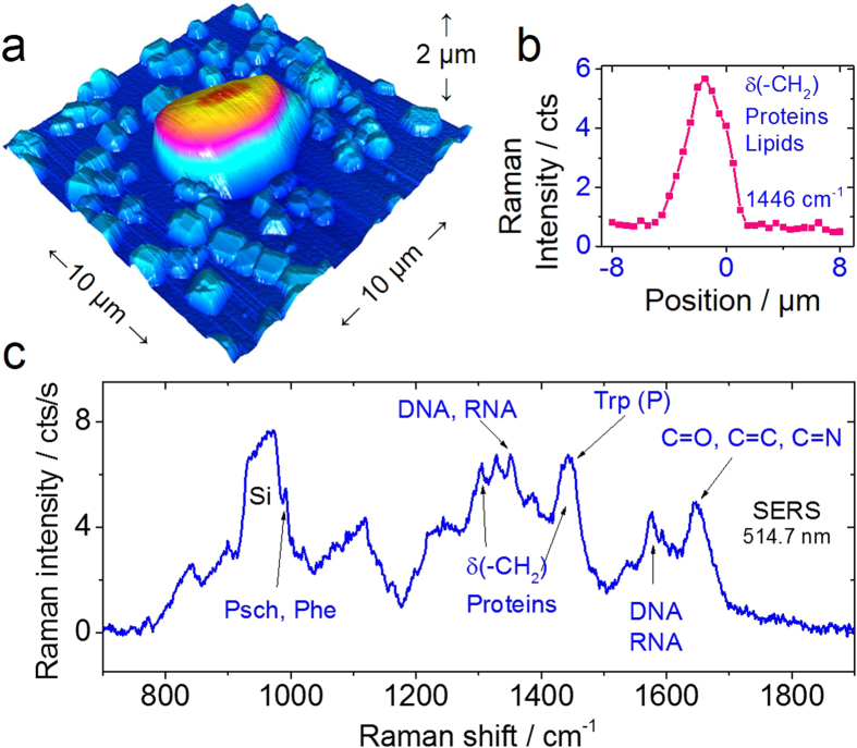
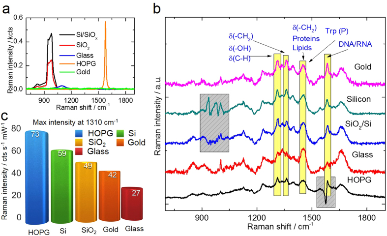
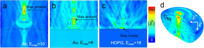
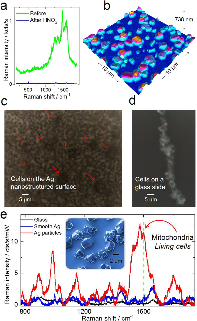
Raman spectroscopy is a powerful analytical method that allows deposited and/or immobilized cells to be evaluated without complex sample preparation or labeling. However, a main limitation of Raman spectroscopy in cell analysis is the extremely weak Raman intensity that results in low signal to noise ratios. Therefore, it is important to seize any opportunity that increases the intensity of the Raman signal and to understand whether and how the signal enhancement changes with respect to the substrate used. Our experimental results show clear differences in the spectroscopic response from cells on different surfaces. This result is partly due to the difference in spatial distribution of electric field at the substrate/cell interface as shown by numerical simulations. We found that the substrate also changes the spatial location of maximum field enhancement around the cells. Moreover, beyond conventional flat surfaces, we introduce an efficient nanostructured silver substrate that largely enhances the Raman signal intensity from a single yeast cell. This work contributes to the field of vibrational spectroscopy analysis by providing a fresh look at the significance of the substrate for Raman investigations in cell research.
拉曼光谱是一种强大的分析方法,它无需复杂的样品制备或标记就能对沉积和/或固定的细胞进行评估。然而,拉曼光谱在细胞分析中的一个主要限制是拉曼强度极其微弱,导致信噪比很低。因此,抓住任何增加拉曼信号强度的机会,并了解信号增强是否以及如何随所用底物而变化,这一点很重要。我们的实验结果表明,不同表面上的细胞在光谱响应上存在明显差异。这一结果部分归因于数值模拟所示的底物/细胞界面处电场空间分布的差异。我们发现,底物还会改变细胞周围最大场增强的空间位置。此外,除了传统的平面表面,我们还引入了一种高效的纳米结构银底物,它能大幅增强单个酵母细胞的拉曼信号强度。这项工作通过重新审视底物在细胞研究拉曼调查中的重要性,为振动光谱分析领域做出了贡献。