Sirenko Oksana, Mitlo Trisha, Hesley Jayne, Luke Steve, Owens Windsor, Cromwell Evan F
1 Molecular Devices , LLC, Sunnyvale, California.
2 Protein Fluidics, Inc. , Palo Alto, California.
Assay Drug Dev Technol. 2015 Sep;13(7):402-14. doi: 10.1089/adt.2015.655.
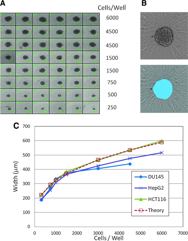
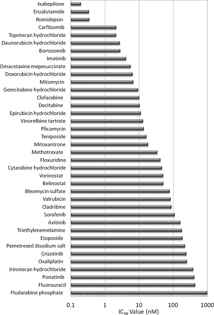
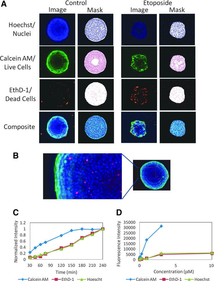
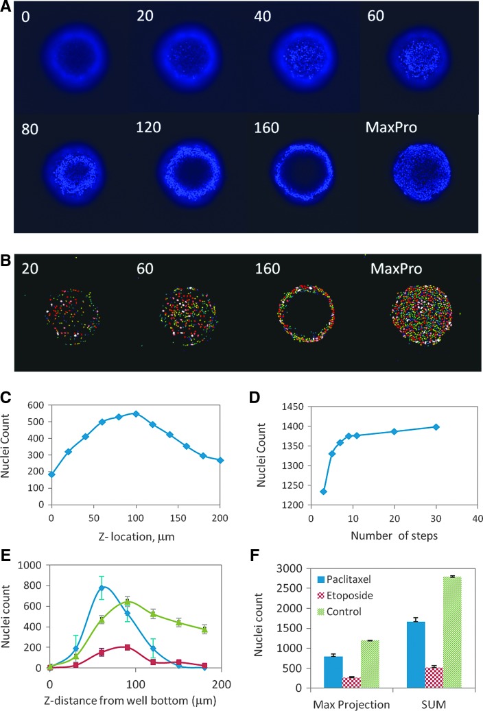
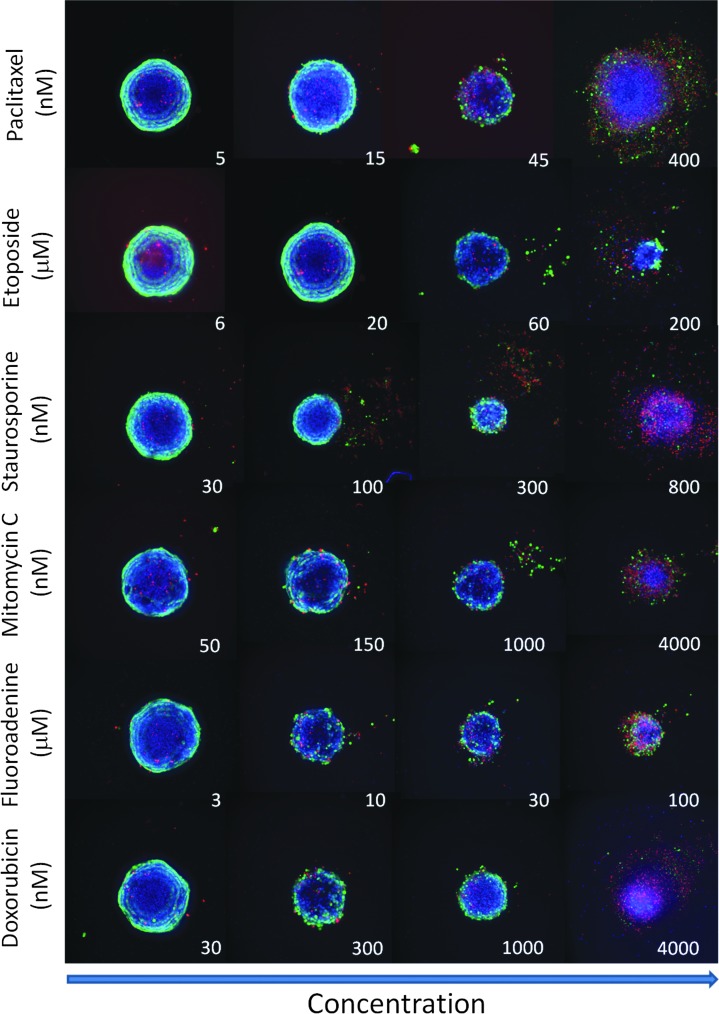
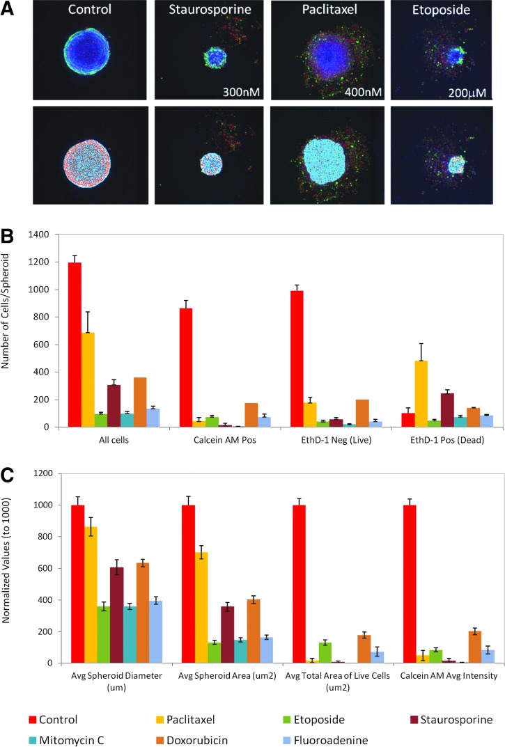
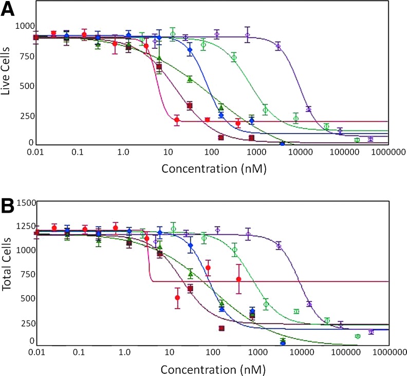
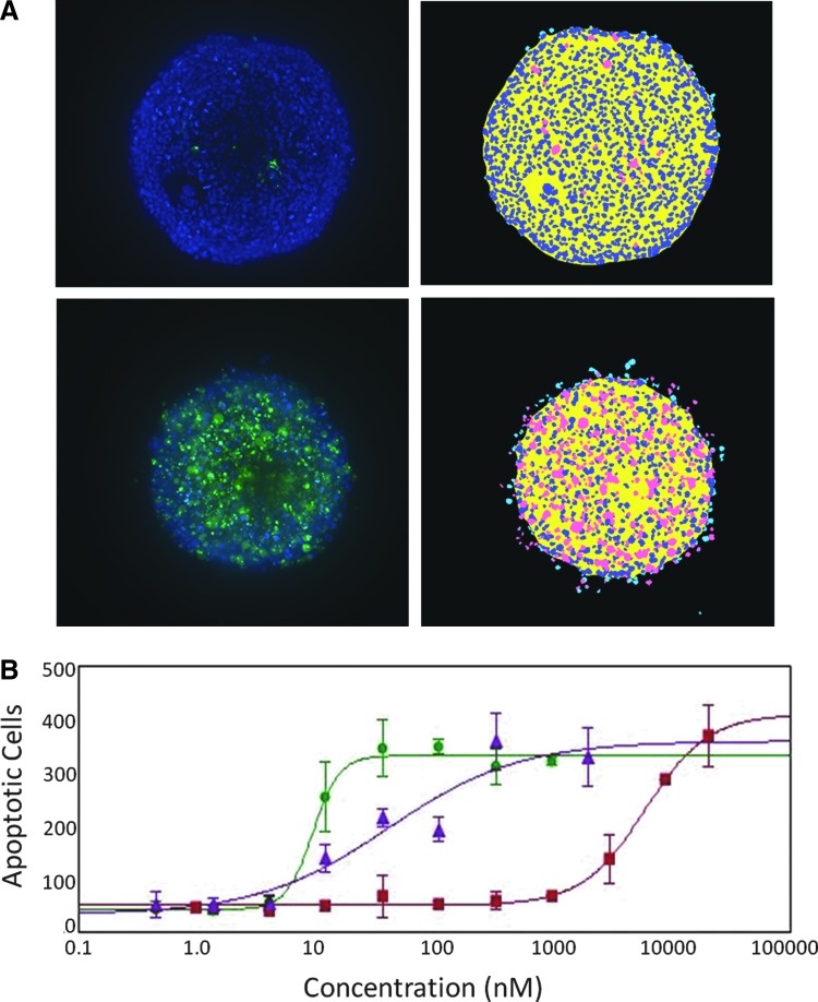
There is an increasing interest in using three-dimensional (3D) spheroids for modeling cancer and tissue biology to accelerate translation research. Development of higher throughput assays to quantify phenotypic changes in spheroids is an active area of investigation. The goal of this study was to develop higher throughput high-content imaging and analysis methods to characterize phenotypic changes in human cancer spheroids in response to compound treatment. We optimized spheroid cell culture protocols using low adhesion U-bottom 96- and 384-well plates for three common cancer cell lines and improved the workflow with a one-step staining procedure that reduces assay time and minimizes variability. We streamlined imaging acquisition by using a maximum projection algorithm that combines cellular information from multiple slices through a 3D object into a single image, enabling efficient comparison of different spheroid phenotypes. A custom image analysis method was implemented to provide multiparametric characterization of single-cell and spheroid phenotypes. We report a number of readouts, including quantification of marker-specific cell numbers, measurement of cell viability and apoptosis, and characterization of spheroid size and shape. Assay performance was assessed using established anticancer cytostatic and cytotoxic drugs. We demonstrated concentration-response effects for different readouts and measured IC50 values, comparing 3D spheroid results to two-dimensional cell cultures. Finally, a library of 119 approved anticancer drugs was screened across a wide range of concentrations using HCT116 colon cancer spheroids. The proposed methods can increase performance and throughput of high-content assays for compound screening and evaluation of anticancer drugs with 3D cell models.
利用三维(3D)球体来模拟癌症和组织生物学以加速转化研究的兴趣与日俱增。开发用于量化球体表型变化的高通量检测方法是一个活跃的研究领域。本研究的目的是开发高通量高内涵成像和分析方法,以表征人癌症球体在化合物处理后的表型变化。我们使用低粘附U型底96孔和384孔板针对三种常见癌细胞系优化了球体细胞培养方案,并通过一步染色程序改进了工作流程,该程序减少了检测时间并使变异性最小化。我们通过使用最大投影算法简化了成像采集,该算法将来自通过3D物体的多个切片的细胞信息组合成单个图像,从而能够有效比较不同的球体表型。实施了一种定制的图像分析方法,以提供单细胞和球体表型的多参数表征。我们报告了许多读数,包括标记物特异性细胞数量的量化、细胞活力和凋亡的测量以及球体大小和形状的表征。使用已确立的抗癌细胞生长抑制剂和细胞毒性药物评估了检测性能。我们展示了不同读数的浓度响应效应并测量了IC50值,将3D球体结果与二维细胞培养进行了比较。最后,使用HCT116结肠癌细胞球体在广泛的浓度范围内筛选了一个包含119种已批准抗癌药物的文库。所提出的方法可以提高使用3D细胞模型进行化合物筛选和抗癌药物评估的高内涵检测的性能和通量。