Loukovaara Sirpa, Gucciardo Erika, Repo Pauliina, Lohi Jouko, Salven Petri, Lehti Kaisa
Unit of Vitreoretinal Surgery, Department of Ophthalmology, Helsinki, Finland.
Genome-Scale Biology Research Program, Research Programs Unit, Biomedicum Helsinki, University of Helsinki, Helsinki, Finland.
Case Rep Ophthalmol. 2015 Jul 17;6(2):228-38. doi: 10.1159/000437254. eCollection 2015 May-Aug.
Pathological vascular differentiation in retinal vein occlusion (RVO)-related neovessel formation remains poorly characterized. The role of intraocular lymphatic-like differentiation or endothelial progenitor cell activity has not been studied in this disease.
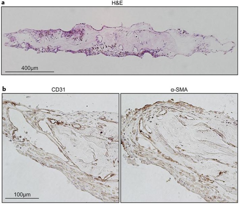
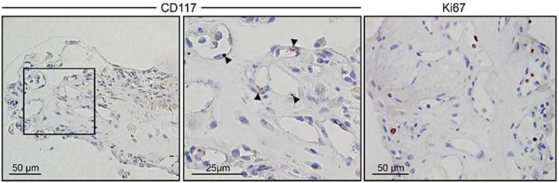
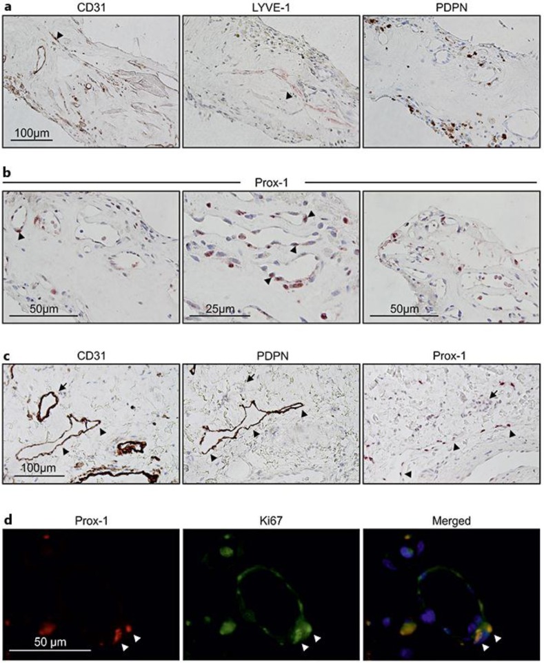
Vitrectomy was performed in an eye with hemi-RVO; the neovessel membrane located at the optic nerve head was removed and subjected to immunohistochemistry. Characterization of the neovascular tissue was performed using hematoxylin and eosin, α-smooth muscle actin, and the pan-endothelial cell (EC) adhesion molecule CD31. The expression of lymphatic EC markers was studied by lymphatic vessel endothelial hyaluronan receptor 1 (LYVE-1), podoplanin (PDPN), and prospero-related homeobox protein 1 (Prox-1). Potential vascular stem/progenitor cells were identified by active cellular proliferation (Ki67) and expression of the stem cell marker CD117.
The specimen contained blood vessels lined by ECs and surrounded by pericytes. Immunoreactivity for LYVE-1 and Prox-1 was detected, with Prox-1 being more widely expressed in the active Ki67-positive lumen-lining cells. PDPN expression was instead found in the cells residing in the extravascular tissue. Expression of the stem cell markers CD117 and Ki67 suggested vascular endothelial progenitor cell activity.
Intraocular lymphatic-like differentiation coupled with progenitor cell activation may be involved in the pathology of neovessel formation in ischemia-induced human hemi-RVO.
视网膜静脉阻塞(RVO)相关新生血管形成中的病理性血管分化仍未得到充分表征。眼内类淋巴管分化或内皮祖细胞活性在该疾病中的作用尚未得到研究。
对一只患有半侧RVO的眼睛进行玻璃体切除术;切除位于视神经乳头的新生血管膜并进行免疫组织化学检查。使用苏木精和伊红、α平滑肌肌动蛋白和全内皮细胞(EC)黏附分子CD31对新生血管组织进行表征。通过淋巴管内皮透明质酸受体1(LYVE-1)、足板蛋白(PDPN)和prospero相关同源盒蛋白1(Prox-1)研究淋巴管内皮标志物的表达。通过活跃的细胞增殖(Ki67)和干细胞标志物CD117的表达鉴定潜在的血管干/祖细胞。
标本中含有由内皮细胞衬里并被周细胞包围的血管。检测到LYVE-1和Prox-1的免疫反应性,其中Prox-1在活跃的Ki67阳性管腔衬里细胞中表达更广泛。相反,在血管外组织中的细胞中发现了PDPN表达。干细胞标志物CD117和Ki67的表达提示血管内皮祖细胞活性。
眼内类淋巴管分化与祖细胞激活可能参与缺血诱导的人类半侧RVO新生血管形成的病理过程。