Lloyd Isaac E, Clement Parker W, Salzman Karen L, Jensen Randy L, Salama Mohamed E, Palmer Cheryl A
Department of Pathology, University of Utah, 15 North Medical Drive East, Suite #1100, Salt Lake City, UT, 84112, USA.
Department of Radiology, University of Utah, 30 N 1900 E, Salt Lake City, UT, 84132, USA.
Diagn Pathol. 2015 Sep 2;10:152. doi: 10.1186/s13000-015-0387-9.
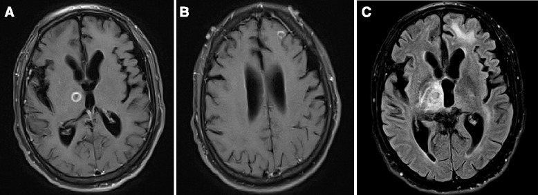
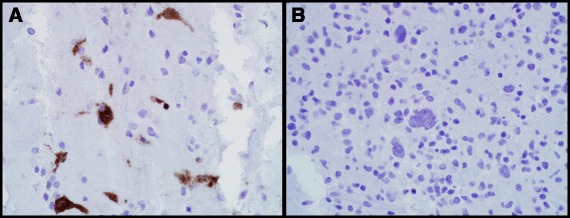
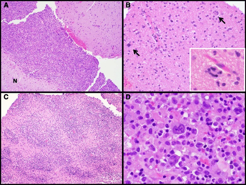
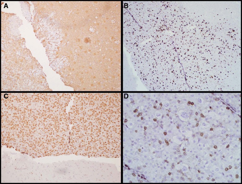
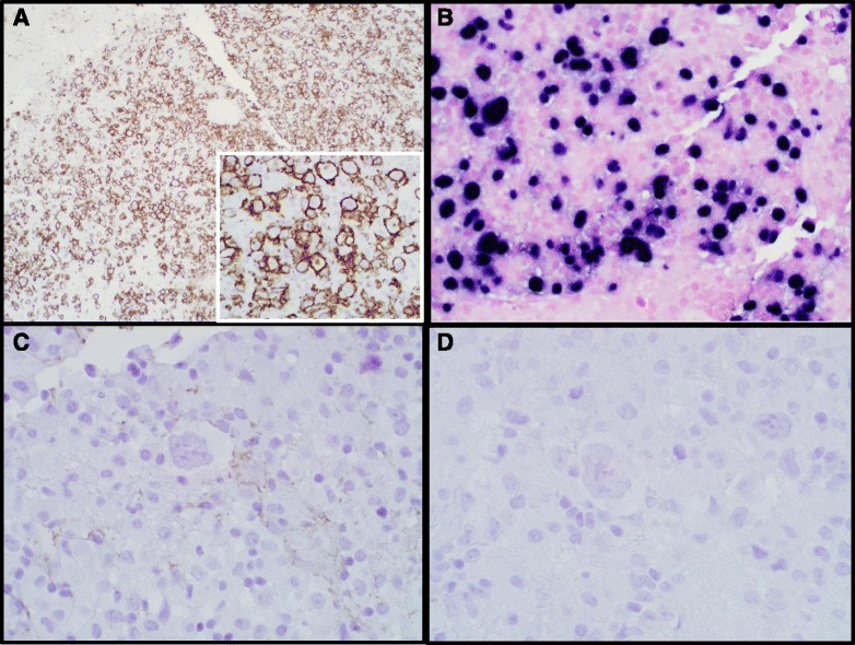
HIV-associated primary CNS lymphomas are well-recognized, almost exclusively EBV-driven neoplasms with poor clinical prognosis. We report a challenging, atypical case of an HIV-associated lymphoproliferative disorder with unusual morphologic features reminiscent of Hodgkin Lymphoma, accompanied by HIV encephalitis. A 52-year-old male presented with acute seizures after seven months of progressive neurocognitive decline that was clinically diagnosed as progressive supranuclear palsy. Clinical work-up revealed HIV infection along with two ring-enhancing lesions in the brain on MRI, and negative CSF EBV testing. Subsequent biopsy showed well-demarcated hypercellular regions in the brain comprised of scattered Reed-Sternberg-like cells in a background of small to medium-sized lymphocytes exhibiting focal angiocentricity and geographic necrosis. The atypical cells were positive for CD20, EBV, and CD79a, and negative for CD45, GFAP, CD15, CD30, and p24. These cells were admixed with numerous CD68-positive cells. The adjacent brain showed classic features of HIV encephalitis with perivascular, CD68 and p24-positive multinucleated giant cells. This case illustrates several diagnostic pitfalls in the work-up of HIV-associated brain lesions, as well as reporting a unique histomorphology for an HIV-related primary CNS lymphoproliferative disorder.
HIV相关的原发性中枢神经系统淋巴瘤是公认的、几乎完全由EB病毒驱动的肿瘤,临床预后较差。我们报告了一例具有挑战性的非典型HIV相关淋巴增殖性疾病病例,其具有不寻常的形态学特征,让人联想到霍奇金淋巴瘤,并伴有HIV脑炎。一名52岁男性在经历了7个月的进行性神经认知衰退后出现急性癫痫发作,临床诊断为进行性核上性麻痹。临床检查发现HIV感染,MRI显示脑部有两个环形强化病变,脑脊液EB病毒检测为阴性。随后的活检显示脑部有界限清楚的细胞增多区域,由散在的里德-斯腾伯格样细胞组成,背景是中小淋巴细胞,表现出局灶性血管中心性和地图状坏死。非典型细胞CD20、EB病毒和CD79a呈阳性,CD45、GFAP、CD15、CD30和p24呈阴性。这些细胞与大量CD-68阳性细胞混合。相邻脑组织显示出HIV脑炎的典型特征,有血管周围、CD68和p24阳性的多核巨细胞。该病例说明了HIV相关脑病变检查中的几个诊断陷阱,同时报告了一种HIV相关原发性中枢神经系统淋巴增殖性疾病独特的组织形态学。