Guo Hongwei, Cheng Yabin, Martinka Magdalena, McElwee Kevin
Department of Dermatology and Skin Science, University of British Columbia, Vancouver, Canada.
Department of Dermatology, Affiliated Hospital of Guangdong Medical College, Zhanjiang, Guangdong, China.
Oncotarget. 2015 Sep 22;6(28):25484-98. doi: 10.18632/oncotarget.4688.
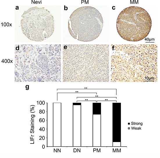
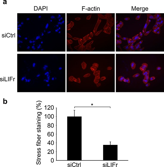
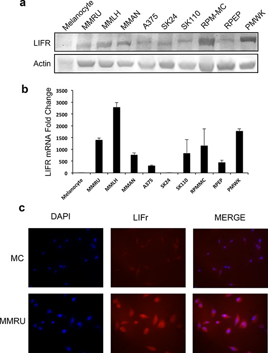
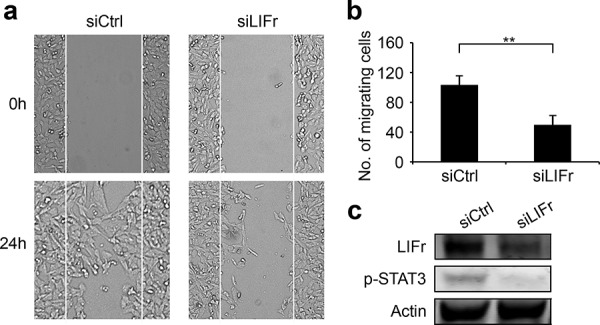
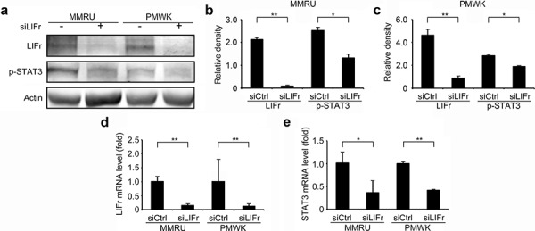
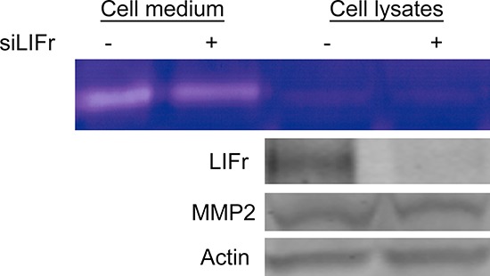
Increased or decreased expression of LIF receptor (LIFr) has been reported in several human cancers, including skin cancer, but its role in melanoma is unknown. In this study, we investigated the expression pattern of LIFr in melanoma and assessed its prognostic value. Using tissue microarrays consisting of 441 melanomas and 96 nevi, we found that no normal nevi showed high LIFr expression. LIFr staining was significantly increased in primary melanoma compared to dysplastic nevi (P = 0.0003) and further increased in metastatic melanoma (P = 0.0000). Kaplan-Meier survival curve and univariate Cox regression analyses showed that increased expression of LIFr was correlated with poorer 5-year patient survival (overall survival, P = 0.0000; disease-specific survival, P = 0.0000). Multivariate Cox regression analyses indicated that increased LIFr expression was an independent prognostic marker for primary melanoma (P = 0.036). LIFr knockdown inhibited melanoma cell migration in wound healing assays and reduced stress fiber formation. LIFr knockdown correlated with STAT3 suppression, but not YAP, suggesting that LIFr activation might stimulate melanoma cell migration through the STAT3 pathway. Our data indicate that strong LIFr expression identifies potentially highly malignant melanocytic lesions at an early stage and LIFr may be a potential target for the development of early intervention therapeutics.
已有报道称,在包括皮肤癌在内的多种人类癌症中,白血病抑制因子受体(LIFr)的表达会升高或降低,但其在黑色素瘤中的作用尚不清楚。在本研究中,我们调查了LIFr在黑色素瘤中的表达模式,并评估了其预后价值。使用由441例黑色素瘤和96例痣组成的组织微阵列,我们发现正常痣均未显示出高LIFr表达。与发育异常痣相比,原发性黑色素瘤中的LIFr染色显著增加(P = 0.0003),而在转移性黑色素瘤中进一步增加(P = 0.0000)。Kaplan-Meier生存曲线和单变量Cox回归分析表明,LIFr表达增加与患者5年生存率较差相关(总生存率,P = 0.0000;疾病特异性生存率,P = 0.0000)。多变量Cox回归分析表明,LIFr表达增加是原发性黑色素瘤的独立预后标志物(P = 0.036)。在伤口愈合试验中,LIFr敲低抑制了黑色素瘤细胞的迁移,并减少了应力纤维的形成。LIFr敲低与STAT3抑制相关,但与YAP无关,这表明LIFr激活可能通过STAT3途径刺激黑色素瘤细胞迁移。我们的数据表明,强烈的LIFr表达可在早期识别潜在的高恶性黑素细胞病变,并且LIFr可能是早期干预治疗开发的潜在靶点。