Ahn Sung Hoon, Oh Tae Hoon, Seo Ill Young
Department of Urology, Institute of Wonkwang Medical Science, Wonkwang University School of Medicine, Iksan, Korea.
Korean J Urol. 2015 Sep;56(9):644-9. doi: 10.4111/kju.2015.56.9.644. Epub 2015 Sep 8.
To assess the potential of dual-energy computed tomography (DECT) to identify urinary stone components, particularly uric acid and calcium oxalate monohydrate, which are unsuitable for extracorporeal shock wave lithotripsy (ESWL).
This clinical study included 246 patients who underwent removal of urinary stones and an analysis of stone components between November 2009 and August 2013. All patients received preoperative DECT using two energy values (80 kVp and 140 kVp). Hounsfield units (HU) were measured and matched to the stone component.
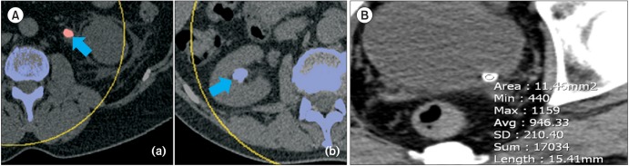
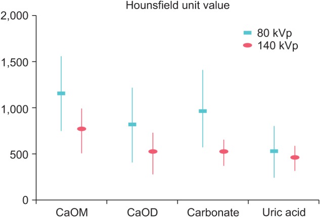
Significant differences in HU values were observed between uric acid and nonuric acid stones at the 80 and 140 kVp energy values (p<0.001). All uric acid stones were red on color-coded DECT images, whereas 96.3% of the nonuric acid stones were blue. Patients with calcium oxalate stones were divided into two groups according to the amount of monohydrate (calcium oxalate monohydrate group: monohydrate≥90%, calcium oxalate dihydrate group: monohydrate<90%). Significant differences in HU values were detected between the two groups at both energy values (p<0.001).
DECT improved the characterization of urinary stone components and was a useful method for identifying uric acid and calcium oxalate monohydrate stones, which are unsuitable for ESWL.
评估双能计算机断层扫描(DECT)识别尿路结石成分的潜力,尤其是尿酸结石和一水草酸钙结石,这两种结石不适合体外冲击波碎石术(ESWL)。
这项临床研究纳入了2009年11月至2013年8月期间接受尿路结石清除及结石成分分析的246例患者。所有患者术前均接受了使用两个能量值(80 kVp和140 kVp)的DECT检查。测量了豪斯菲尔德单位(HU)并将其与结石成分进行匹配。
在80 kVp和140 kVp能量值下,尿酸结石和非尿酸结石的HU值存在显著差异(p<0.001)。在彩色编码的DECT图像上,所有尿酸结石均为红色,而96.3%的非尿酸结石为蓝色。根据一水草酸钙的含量,草酸钙结石患者被分为两组(一水草酸钙组:一水草酸钙≥90%,二水草酸钙组:一水草酸钙<90%)。在两个能量值下,两组之间的HU值均存在显著差异(p<0.001)。
DECT改善了尿路结石成分的特征描述,是识别不适合ESWL的尿酸结石和一水草酸钙结石的有用方法。