• 文献检索
  • 文档翻译
  • 深度研究
  • 学术资讯
  • Suppr Zotero 插件Zotero 插件
  • 邀请有礼
  • 套餐&价格
  • 历史记录
应用&插件
Suppr Zotero 插件Zotero 插件浏览器插件Mac 客户端Windows 客户端微信小程序
定价
高级版会员购买积分包购买API积分包
服务
文献检索文档翻译深度研究API 文档MCP 服务
关于我们
关于 Suppr公司介绍联系我们用户协议隐私条款
关注我们

Suppr 超能文献

核心技术专利:CN118964589B侵权必究
粤ICP备2023148730 号-1Suppr @ 2026

文献检索

告别复杂PubMed语法,用中文像聊天一样搜索,搜遍4000万医学文献。AI智能推荐,让科研检索更轻松。

立即免费搜索

文件翻译

保留排版,准确专业,支持PDF/Word/PPT等文件格式,支持 12+语言互译。

免费翻译文档

深度研究

AI帮你快速写综述,25分钟生成高质量综述,智能提取关键信息,辅助科研写作。

立即免费体验

从牙科CT图像中快速准确地半自动分割单个牙齿

Fast and Accurate Semiautomatic Segmentation of Individual Teeth from Dental CT Images.

作者信息

Kang Ho Chul, Choi Chankyu, Shin Juneseuk, Lee Jeongjin, Shin Yeong-Gil

机构信息

Palette Soft Inc., 599 Kwanak-ro, Kwanak-gu, Seoul 151-742, Republic of Korea.

Planet SK Planet Co., Ltd., Bundang-gu, 264 Pangyo-ro, Seongnam-si, Gyeonggi-do 463-400, Republic of Korea.

出版信息

Comput Math Methods Med. 2015;2015:810796. doi: 10.1155/2015/810796. Epub 2015 Aug 27.

DOI:10.1155/2015/810796
PMID:26413143
原文链接:https://pmc.ncbi.nlm.nih.gov/articles/PMC4564792/
Abstract

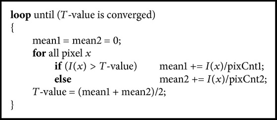
In this paper, we propose a fast and accurate semiautomatic method to effectively distinguish individual teeth from the sockets of teeth in dental CT images. Parameter values of thresholding and shapes of the teeth are propagated to the neighboring slice, based on the separated teeth from reference images. After the propagation of threshold values and shapes of the teeth, the histogram of the current slice was analyzed. The individual teeth are automatically separated and segmented by using seeded region growing. Then, the newly generated separation information is iteratively propagated to the neighboring slice. Our method was validated by ten sets of dental CT scans, and the results were compared with the manually segmented result and conventional methods. The average error of absolute value of volume measurement was 2.29 ± 0.56%, which was more accurate than conventional methods. Boosting up the speed with the multicore processors was shown to be 2.4 times faster than a single core processor. The proposed method identified the individual teeth accurately, demonstrating that it can give dentists substantial assistance during dental surgery.

摘要

在本文中,我们提出了一种快速且准确的半自动方法,以有效区分牙科CT图像中牙齿与牙槽窝中的单个牙齿。基于参考图像中分离出的牙齿,阈值参数值和牙齿形状会传播到相邻切片。在牙齿阈值和形状传播之后,对当前切片的直方图进行分析。通过使用种子区域生长法自动分离并分割单个牙齿。然后,将新生成的分离信息迭代传播到相邻切片。我们的方法通过十组牙科CT扫描进行了验证,并将结果与手动分割结果和传统方法进行了比较。体积测量绝对值的平均误差为2.29±0.56%,比传统方法更准确。使用多核处理器提高速度后,显示比单核处理器快2.4倍。所提出的方法能够准确识别单个牙齿,表明它可以在牙科手术期间为牙医提供实质性帮助。

https://cdn.ncbi.nlm.nih.gov/pmc/blobs/1576/4564792/3b97756b474a/CMMM2015-810796.pseudo.002.jpg
https://cdn.ncbi.nlm.nih.gov/pmc/blobs/1576/4564792/87758b1e69a8/CMMM2015-810796.001.jpg
https://cdn.ncbi.nlm.nih.gov/pmc/blobs/1576/4564792/ef2820751fae/CMMM2015-810796.002.jpg
https://cdn.ncbi.nlm.nih.gov/pmc/blobs/1576/4564792/60ddb0b8fdeb/CMMM2015-810796.003.jpg
https://cdn.ncbi.nlm.nih.gov/pmc/blobs/1576/4564792/456e1126873d/CMMM2015-810796.004.jpg
https://cdn.ncbi.nlm.nih.gov/pmc/blobs/1576/4564792/50f80e6f6bcb/CMMM2015-810796.005.jpg
https://cdn.ncbi.nlm.nih.gov/pmc/blobs/1576/4564792/b2fcd50f2470/CMMM2015-810796.006.jpg
https://cdn.ncbi.nlm.nih.gov/pmc/blobs/1576/4564792/3608d0f4de53/CMMM2015-810796.007.jpg
https://cdn.ncbi.nlm.nih.gov/pmc/blobs/1576/4564792/4dc61680a6dd/CMMM2015-810796.008.jpg
https://cdn.ncbi.nlm.nih.gov/pmc/blobs/1576/4564792/e9e1065f3e11/CMMM2015-810796.009.jpg
https://cdn.ncbi.nlm.nih.gov/pmc/blobs/1576/4564792/3934b619d298/CMMM2015-810796.010.jpg
https://cdn.ncbi.nlm.nih.gov/pmc/blobs/1576/4564792/c6b53804399b/CMMM2015-810796.011.jpg
https://cdn.ncbi.nlm.nih.gov/pmc/blobs/1576/4564792/f6e5adaf47af/CMMM2015-810796.012.jpg
https://cdn.ncbi.nlm.nih.gov/pmc/blobs/1576/4564792/6bcbda296336/CMMM2015-810796.013.jpg
https://cdn.ncbi.nlm.nih.gov/pmc/blobs/1576/4564792/6440eeccf22c/CMMM2015-810796.014.jpg
https://cdn.ncbi.nlm.nih.gov/pmc/blobs/1576/4564792/e7bda751a787/CMMM2015-810796.pseudo.001.jpg
https://cdn.ncbi.nlm.nih.gov/pmc/blobs/1576/4564792/3b97756b474a/CMMM2015-810796.pseudo.002.jpg

相似文献

1
Fast and Accurate Semiautomatic Segmentation of Individual Teeth from Dental CT Images.从牙科CT图像中快速准确地半自动分割单个牙齿
Comput Math Methods Med. 2015;2015:810796. doi: 10.1155/2015/810796. Epub 2015 Aug 27.
2
Automatic teeth axes calculation for well-aligned teeth using cost profile analysis along teeth center arch.利用沿牙齿中心弓的成本剖面分析计算排列整齐的牙齿的自动牙齿轴。
IEEE Trans Biomed Eng. 2012 Apr;59(4):1145-54. doi: 10.1109/TBME.2012.2185825. Epub 2012 Jan 24.
3
Individual tooth segmentation from CT images scanned with contacts of maxillary and mandible teeth.从上颌和下颌牙齿接触扫描的 CT 图像中进行单个牙齿分割。
Comput Methods Programs Biomed. 2017 Jan;138:1-12. doi: 10.1016/j.cmpb.2016.10.002. Epub 2016 Oct 14.
4
[A semi-automatic method for tooth segmentation in dental CT images].[一种用于牙科CT图像中牙齿分割的半自动方法]
Sheng Wu Yi Xue Gong Cheng Xue Za Zhi. 2007 Feb;24(1):15-8.
5
Tooth and Alveolar Bone Segmentation From Dental Computed Tomography Images.从牙科计算机断层扫描图像中进行牙齿和牙槽骨分割。
IEEE J Biomed Health Inform. 2018 Jan;22(1):196-204. doi: 10.1109/JBHI.2017.2709406. Epub 2017 May 29.
6
Toward accurate tooth segmentation from computed tomography images using a hybrid level set model.使用混合水平集模型从计算机断层扫描图像中实现准确的牙齿分割。
Med Phys. 2015 Jan;42(1):14-27. doi: 10.1118/1.4901521.
7
[Observations on three dimensional images for cracks of doweled teeth--comparison of images from specimen sections and dental tomograms from small three dimensional X-ray CT].关于桩核牙裂纹三维图像的观察——标本切片图像与小型三维X射线CT牙科断层扫描图像的比较
Nihon Hotetsu Shika Gakkai Zasshi. 2005 Feb;49(1):84-92. doi: 10.2186/jjps.49.84.
8
[Quantitative Segmentation and Measurement of Tooth from Computed Tomography Image Based on Regional Adaptive Deformation Model].基于区域自适应变形模型的计算机断层扫描图像中牙齿的定量分割与测量
Sheng Wu Yi Xue Gong Cheng Xue Za Zhi. 2016 Apr;33(2):308-14.
9
Tooth shape reconstruction from dental CT images with the region-growing method.基于区域生长法的牙体 CT 图像的牙齿形态重建。
Dentomaxillofac Radiol. 2014;43(6):20140080. doi: 10.1259/dmfr.20140080. Epub 2014 May 2.
10
Computation of tooth axes of existent and missing teeth from 3D CT images.
Biomed Tech (Berl). 2015 Dec;60(6):623-32. doi: 10.1515/bmt-2014-0111.

引用本文的文献

1
Semi or fully automatic tooth segmentation in CBCT images: a review.锥形束计算机断层扫描(CBCT)图像中的半自动或全自动牙齿分割:综述
PeerJ Comput Sci. 2024 Apr 19;10:e1994. doi: 10.7717/peerj-cs.1994. eCollection 2024.
2
Improving performance of deep learning models using 3.5D U-Net via majority voting for tooth segmentation on cone beam computed tomography.使用 3.5D U-Net 通过多数投票提高基于锥形束计算机断层扫描的牙齿分割的深度学习模型性能。
Sci Rep. 2022 Nov 17;12(1):19809. doi: 10.1038/s41598-022-23901-7.
3
Refined tooth and pulp segmentation using U-Net in CBCT image.

本文引用的文献

1
Segmentation of Trabecular Jaw Bone on Cone Beam CT Datasets.锥形束CT数据集上的下颌骨小梁分割
Clin Implant Dent Relat Res. 2015 Dec;17(6):1082-91. doi: 10.1111/cid.12217. Epub 2014 Mar 14.
2
Interactions between magnetic resonance imaging and dental material.磁共振成像与牙科材料之间的相互作用。
J Pharm Bioallied Sci. 2013 Jun;5(Suppl 1):S113-6. doi: 10.4103/0975-7406.113309.
3
Automatic teeth axes calculation for well-aligned teeth using cost profile analysis along teeth center arch.利用沿牙齿中心弓的成本剖面分析计算排列整齐的牙齿的自动牙齿轴。
基于 CBCT 图像的 U-Net 进行精细化牙及牙髓分割。
Dentomaxillofac Radiol. 2021 Sep 1;50(6):20200251. doi: 10.1259/dmfr.20200251. Epub 2021 Jan 15.
4
Three-dimensional evaluation of morphology and position of impacted supernumerary teeth in cases of cleidocranial dysplasia.髁颅发育不全病例中埋伏多生牙的形态和位置的三维评估。
Congenit Anom (Kyoto). 2020 Jul;60(4):106-114. doi: 10.1111/cga.12358. Epub 2019 Oct 23.
IEEE Trans Biomed Eng. 2012 Apr;59(4):1145-54. doi: 10.1109/TBME.2012.2185825. Epub 2012 Jan 24.
4
Automatic extraction of inferior alveolar nerve canal using feature-enhancing panoramic volume rendering.使用特征增强全景容积渲染自动提取下颌神经管。
IEEE Trans Biomed Eng. 2011 Feb;58(2):253-64. doi: 10.1109/TBME.2010.2089053.
5
Virtual dental surgery as a new educational tool in dental school.虚拟牙科手术作为牙科学院的一种新的教育工具。
J Craniomaxillofac Surg. 2010 Dec;38(8):560-4. doi: 10.1016/j.jcms.2010.02.011. Epub 2010 Mar 29.
6
Three-dimensional treatment planning of orthognathic surgery in the era of virtual imaging.虚拟成像时代正颌外科的三维治疗计划
J Oral Maxillofac Surg. 2009 Oct;67(10):2080-92. doi: 10.1016/j.joms.2009.06.007.
7
A cone-beam computed tomography triple scan procedure to obtain a three-dimensional augmented virtual skull model appropriate for orthognathic surgery planning.一种用于获取适用于正颌外科手术规划的三维增强虚拟颅骨模型的锥形束计算机断层扫描三重扫描程序。
J Craniofac Surg. 2009 Mar;20(2):297-307. doi: 10.1097/SCS.0b013e3181996803.
8
Morphometric analysis of white matter lesions in MR images: method and validation.磁共振图像中脑白质病变的形态计量分析:方法与验证。
IEEE Trans Med Imaging. 1994;13(4):716-24. doi: 10.1109/42.363096.
9
Automatic segmentation of jaw tissues in CT using active appearance models and semi-automatic landmarking.使用主动外观模型和半自动地标法在CT中自动分割颌骨组织
Med Image Comput Comput Assist Interv. 2006;9(Pt 1):167-74. doi: 10.1007/11866565_21.
10
Computer methods for automating preoperative dental implant planning: implant positioning and size assignment.用于术前牙种植规划自动化的计算机方法:种植体定位与尺寸确定
Comput Methods Programs Biomed. 2007 Apr;86(1):30-8. doi: 10.1016/j.cmpb.2006.12.010. Epub 2007 Jan 30.