Lin Ling, Chen Yong-Song, Yao Yan-Dan, Chen Jing-Qi, Chen Jia-Ning, Huang Song-Yin, Zeng Yun-Jie, Yao He-Rui, Zeng Si-Hai, Fu Yong-Shui, Song Er-Wei
Guangdong Provincial Key Laboratory of Malignant Tumor Epigenetics and Gene Regulation, Medical Research Center, Sun Yat-Sen Memorial Hospital, Sun Yat-Sen University, Guangzhou 510120, P. R. China.
Breast Tumor Center, Sun Yat-Sen Memorial Hospital, Sun Yat-Sen University, Guangzhou 510120, P. R. China.
Oncotarget. 2015 Oct 27;6(33):34758-73. doi: 10.18632/oncotarget.5325.
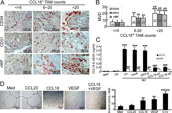
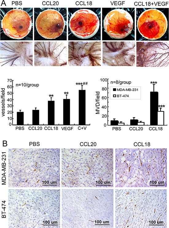
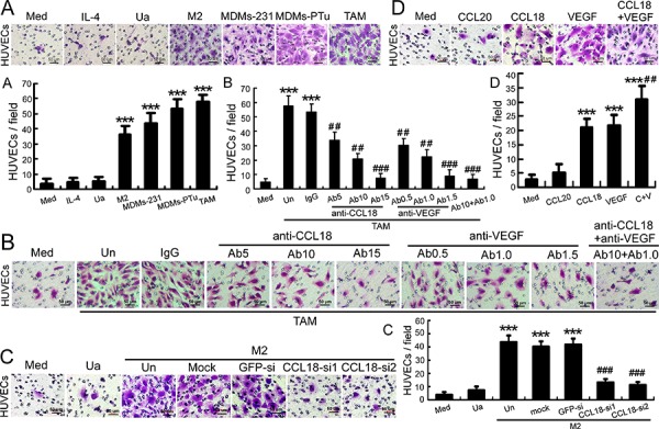
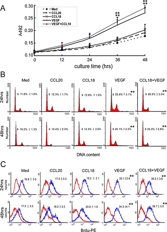
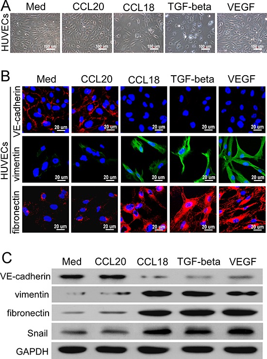
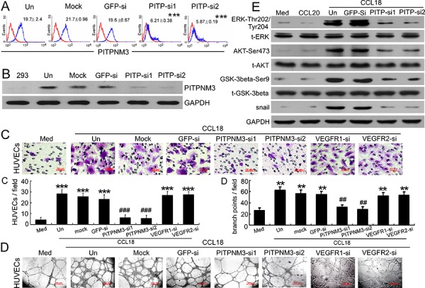
The infiltration of tumor-associated macrophages (TAMs) is associated with extensive angiogenesis, which contributes to a poor prognosis in breast cancer. However, anti-angiogenic therapy with VEGF-specific monotherapy has been unsuccessful in treating breast cancer, and the molecular mechanisms associated with chemoresistance remain unclear. Here, we investigated whether CCL18, a chemokine produced by TAMs, can stimulate angiogenesis in breast cancer, as well as the underlying mechanisms. Double immunohistochemical staining for CCL18 and CD34/CD31/vWF was performed in 80 breast cancer samples to study the correlation between CCL18+ TAMs and microvascular density (MVD). Cocultures of TAMs with human umbilical vein endothelial cells (HUVECs) were used to model the inflammatory microenvironment, and CCL18-induced angiogenesis was evaluated both in vitro and in vivo. We demonstrated that CCL18+ TAM infiltration positively associated with MVD in breast cancer samples, which was correlated with tumor metastasis and poor prognosis. We confirmed, both in vitro and in vivo, that CCL18 and VEGF synergistically promoted endothelial cell migration and angiogenesis. Conversely, blocking CCL18 or VEGF with neutralizing antibodies synergistically inhibited the promigratory effects of TAMs. Silencing PITPNM3, a putative CCL18 receptor, on the surface of HUVECs abrogated CCL18-mediated promigration and the enhancement of HUVEC tube formation, independently of VEGFR signaling. Moreover, CCL18 exposure induced the endothelial-mesenchymal transformation and activated ERK and Akt/GSK-3β/Snail signaling in HUVECs, thereby contributing to its pro-angiogenic effects. In conclusion, our findings suggest that CCL18 released from TAMs promotes angiogenesis and tumor progression in breast cancer; thus, CCL18 may serve as a novel target for anti-angiogenic therapies.
肿瘤相关巨噬细胞(TAM)的浸润与广泛的血管生成相关,这导致乳腺癌预后不良。然而,VEGF特异性单一疗法的抗血管生成治疗在治疗乳腺癌方面并不成功,且与化疗耐药相关的分子机制仍不清楚。在此,我们研究了TAM产生的趋化因子CCL18是否能刺激乳腺癌中的血管生成及其潜在机制。对80例乳腺癌样本进行CCL18与CD34/CD31/vWF的双重免疫组化染色,以研究CCL18+ TAM与微血管密度(MVD)之间的相关性。将TAM与人脐静脉内皮细胞(HUVEC)共培养以模拟炎症微环境,并在体外和体内评估CCL18诱导的血管生成。我们证明,CCL18+ TAM浸润与乳腺癌样本中的MVD呈正相关,这与肿瘤转移和不良预后相关。我们在体外和体内均证实,CCL18和VEGF协同促进内皮细胞迁移和血管生成。相反,用中和抗体阻断CCL18或VEGF可协同抑制TAM的促迁移作用。沉默HUVEC表面假定的CCL18受体PITPNM3可消除CCL18介导的促迁移作用以及HUVEC管形成的增强,且不依赖于VEGFR信号传导。此外,CCL18暴露诱导HUVEC发生内皮-间充质转化并激活ERK和Akt/GSK-3β/Snail信号传导,从而促进其促血管生成作用。总之,我们的研究结果表明,TAM释放的CCL18促进乳腺癌中的血管生成和肿瘤进展;因此,CCL18可能作为抗血管生成治疗的新靶点。