• 文献检索
  • 文档翻译
  • 深度研究
  • 学术资讯
  • Suppr Zotero 插件Zotero 插件
  • 邀请有礼
  • 套餐&价格
  • 历史记录
应用&插件
Suppr Zotero 插件Zotero 插件浏览器插件Mac 客户端Windows 客户端微信小程序
定价
高级版会员购买积分包购买API积分包
服务
文献检索文档翻译深度研究API 文档MCP 服务
关于我们
关于 Suppr公司介绍联系我们用户协议隐私条款
关注我们

Suppr 超能文献

核心技术专利:CN118964589B侵权必究
粤ICP备2023148730 号-1Suppr @ 2026

文献检索

告别复杂PubMed语法,用中文像聊天一样搜索,搜遍4000万医学文献。AI智能推荐,让科研检索更轻松。

立即免费搜索

文件翻译

保留排版,准确专业,支持PDF/Word/PPT等文件格式,支持 12+语言互译。

免费翻译文档

深度研究

AI帮你快速写综述,25分钟生成高质量综述,智能提取关键信息,辅助科研写作。

立即免费体验

使用四维相位对比磁共振成像对胸主动脉内的涡流进行定量分析。

Quantitative Analysis of Vortical Blood Flow in the Thoracic Aorta Using 4D Phase Contrast MRI.

作者信息

von Spiczak Jochen, Crelier Gerard, Giese Daniel, Kozerke Sebastian, Maintz David, Bunck Alexander Christian

机构信息

Department of Radiology and Neuroradiology, University Hospital of Cologne, Cologne, Germany.

Institute for Biomedical Engineering, University and ETH Zurich, Zurich, Switzerland.

出版信息

PLoS One. 2015 Sep 29;10(9):e0139025. doi: 10.1371/journal.pone.0139025. eCollection 2015.

DOI:10.1371/journal.pone.0139025
PMID:26418327
原文链接:https://pmc.ncbi.nlm.nih.gov/articles/PMC4587936/
Abstract

INTRODUCTION

Phase contrast MRI allows for the examination of complex hemodynamics in the heart and adjacent great vessels. Vortex flow patterns seem to play an important role in certain vascular pathologies. We propose two- and three-dimensional metrics for the objective quantification of aortic vortex blood flow in 4D phase contrast MRI.

MATERIALS AND METHODS

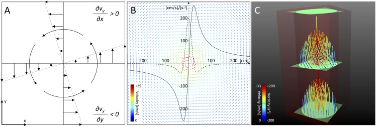
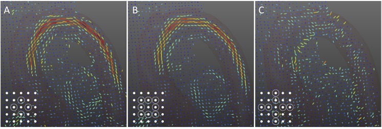
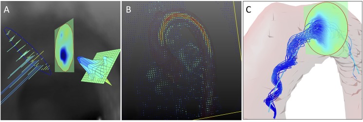
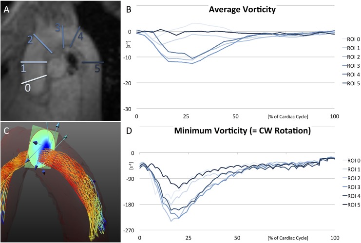
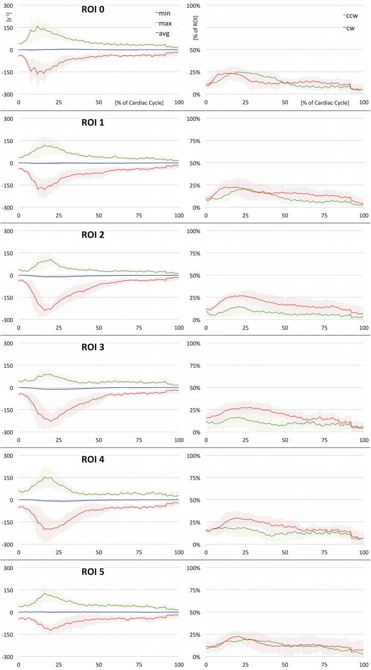
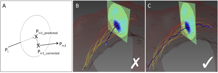
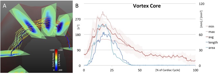
For two-dimensional vorticity assessment, a standardized set of 6 regions-of-interest (ROIs) was defined throughout the course of the aorta. For each ROI, a heatmap of time-resolved vorticity values [Formula: see text] was computed. Evolution of minimum, maximum, and average values as well as opposing rotational flow components were analyzed. For three-dimensional analysis, vortex core detection was implemented combining the predictor-corrector method with λ2 correction. Strength, elongation, and radial expansion of the detected vortex core were recorded over time. All methods were applied to 4D flow MRI datasets of 9 healthy subjects, 2 patients with mildly dilated aorta, and 1 patient with aortic aneurysm.

RESULTS

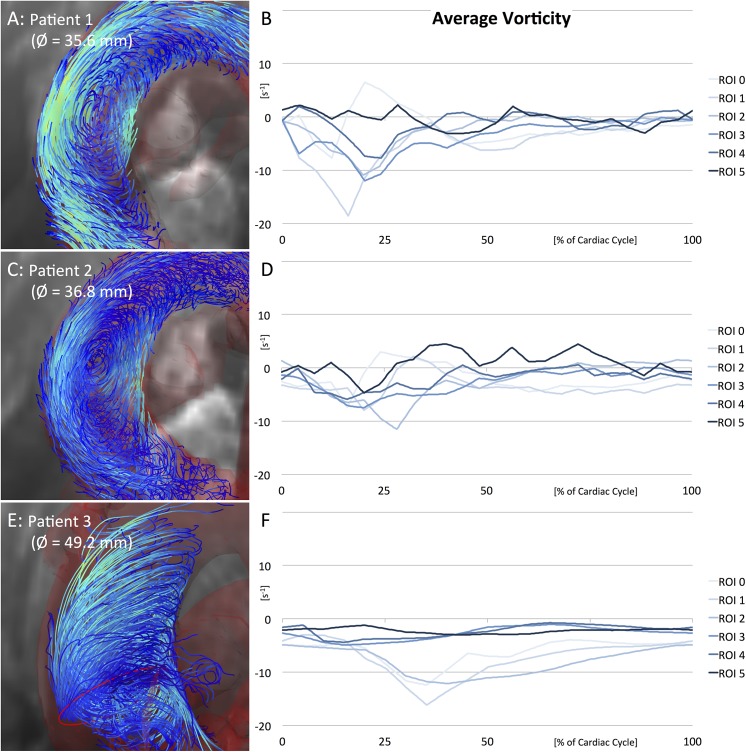
Vorticity quantification in the 6 standardized ROIs enabled the description of physiological vortex flow in the healthy aorta. Helical flow developed early in the ascending aorta (absolute vorticity = 166.4±86.4 s-1 at 12% of cardiac cycle) followed by maximum values in mid-systole in the aortic arch (240.1±45.2 s-1 at 16%). Strength, elongation, and radial expansion of 3D vortex cores escalated in early systole, reaching a peak in mid systole (strength = 241.2±30.7 s-1 at 17%, elongation = 65.1±34.6 mm at 18%, expansion = 80.1±48.8 mm2 at 20%), before all three parameters similarly decreased to overall low values in diastole. Flow patterns were considerably altered in patient data: Vortex flow developed late in mid/end-systole close to the aortic bulb and no physiological helix was found in the aortic arch.

CONCLUSIONS

We have introduced objective measures for quantification of vortical flow in 4D phase contrast MRI. Vortex blood flow in the thoracic aorta could be consistently described in all healthy volunteers. In patient data, pathologically altered vortex flow was observed.

摘要

引言

相位对比磁共振成像(MRI)可用于检查心脏及相邻大血管中的复杂血流动力学。涡流模式似乎在某些血管病变中起重要作用。我们提出了二维和三维指标,用于在四维相位对比MRI中对主动脉涡流血流进行客观量化。

材料与方法

对于二维涡度评估,在整个主动脉行程中定义了一组标准化的6个感兴趣区域(ROI)。对于每个ROI,计算时间分辨涡度值的热图[公式:见原文]。分析最小值、最大值和平均值的演变以及相反的旋转流分量。对于三维分析,结合预测校正法和λ2校正实现涡核检测。记录检测到的涡核的强度、伸长率和径向扩展随时间的变化。所有方法均应用于9名健康受试者、2名轻度主动脉扩张患者和1名主动脉瘤患者的四维血流MRI数据集。

结果

在6个标准化ROI中进行涡度量化能够描述健康主动脉中的生理性涡流。螺旋流在升主动脉早期形成(心动周期的12%时绝对涡度=166.4±86.4 s-1),随后在主动脉弓收缩中期达到最大值(16%时为240.1±45.2 s-1)。三维涡核的强度、伸长率和径向扩展在收缩早期升高,在收缩中期达到峰值(强度=17%时为241.2±30.7 s-1,伸长率=18%时为65.1±34.6 mm,扩展=20%时为80.1±48.8 mm2),然后这三个参数在舒张期均类似地降至总体低值。患者数据中的血流模式有显著改变:涡流在收缩中期/末期接近主动脉球处形成较晚,且在主动脉弓中未发现生理性螺旋。

结论

我们引入了在四维相位对比MRI中量化涡流的客观测量方法。在所有健康志愿者中均可一致地描述胸主动脉中的涡流血流。在患者数据中,观察到了病理性改变的涡流。

https://cdn.ncbi.nlm.nih.gov/pmc/blobs/7073/4587936/62621e8ff185/pone.0139025.g008.jpg
https://cdn.ncbi.nlm.nih.gov/pmc/blobs/7073/4587936/be0a69801f85/pone.0139025.g001.jpg
https://cdn.ncbi.nlm.nih.gov/pmc/blobs/7073/4587936/21ab8077b337/pone.0139025.g002.jpg
https://cdn.ncbi.nlm.nih.gov/pmc/blobs/7073/4587936/cdc6216b9e95/pone.0139025.g003.jpg
https://cdn.ncbi.nlm.nih.gov/pmc/blobs/7073/4587936/8727c07c9095/pone.0139025.g004.jpg
https://cdn.ncbi.nlm.nih.gov/pmc/blobs/7073/4587936/38b7dc485263/pone.0139025.g005.jpg
https://cdn.ncbi.nlm.nih.gov/pmc/blobs/7073/4587936/d78d7bb7fc4b/pone.0139025.g006.jpg
https://cdn.ncbi.nlm.nih.gov/pmc/blobs/7073/4587936/b8d3acf73d16/pone.0139025.g007.jpg
https://cdn.ncbi.nlm.nih.gov/pmc/blobs/7073/4587936/62621e8ff185/pone.0139025.g008.jpg

相似文献

1
Quantitative Analysis of Vortical Blood Flow in the Thoracic Aorta Using 4D Phase Contrast MRI.使用四维相位对比磁共振成像对胸主动脉内的涡流进行定量分析。
PLoS One. 2015 Sep 29;10(9):e0139025. doi: 10.1371/journal.pone.0139025. eCollection 2015.
2
Flow-sensitive four-dimensional magnetic resonance imaging: flow patterns in ascending aortic aneurysms.血流敏感型四维磁共振成像:升主动脉瘤中的血流模式
Eur J Cardiothorac Surg. 2008 Jul;34(1):11-6. doi: 10.1016/j.ejcts.2008.03.047. Epub 2008 Jun 2.
3
Preclinical 4D-flow magnetic resonance phase contrast imaging of the murine aortic arch.小鼠主动脉弓的临床前四维血流磁共振相位对比成像
PLoS One. 2017 Nov 8;12(11):e0187596. doi: 10.1371/journal.pone.0187596. eCollection 2017.
4
Flow-sensitive 4D MRI of the thoracic aorta: comparison of image quality, quantitative flow, and wall parameters at 1.5 T and 3 T.胸主动脉的流量敏感 4D MRI:1.5T 和 3T 时图像质量、定量流量和管壁参数的比较。
J Magn Reson Imaging. 2012 Nov;36(5):1097-103. doi: 10.1002/jmri.23735. Epub 2012 Jun 28.
5
Time-resolved 3-dimensional magnetic resonance phase contrast imaging (4D Flow MRI) reveals altered blood flow patterns in the ascending aorta of patients with valve-sparing aortic root replacement.时分辨 3 维磁共振相位对比成像(4DFlowMRI)显示,在保留主动脉瓣的主动脉根部置换术后患者的升主动脉中,血流模式发生改变。
J Thorac Cardiovasc Surg. 2020 Mar;159(3):798-810.e1. doi: 10.1016/j.jtcvs.2019.02.127. Epub 2019 Apr 5.
6
Imaging of the thoracic aorta with time-resolved three-dimensional phase-contrast MRI: a review.时间分辨三维相位对比磁共振成像对胸主动脉的成像:综述
Semin Thorac Cardiovasc Surg. 2008 Winter;20(4):358-64. doi: 10.1053/j.semtcvs.2008.11.013.
7
Evaluation of 3D blood flow patterns and wall shear stress in the normal and dilated thoracic aorta using flow-sensitive 4D CMR.应用 4D 血流敏感磁共振成像评估正常和扩张性胸主动脉的三维血流模式和壁面切应力。
J Cardiovasc Magn Reson. 2012 Dec 13;14(1):84. doi: 10.1186/1532-429X-14-84.
8
Comparison of 4D flow and 2D velocity-encoded phase contrast MRI sequences for the evaluation of aortic hemodynamics.用于评估主动脉血流动力学的4D流与2D速度编码相位对比MRI序列的比较。
Int J Cardiovasc Imaging. 2016 Oct;32(10):1529-41. doi: 10.1007/s10554-016-0938-5. Epub 2016 Jul 19.
9
Evaluating a Phase-Specific Approach to Aortic Flow: A 4D Flow MRI Study.评估主动脉血流的阶段性方法:一项 4D 流 MRI 研究。
J Magn Reson Imaging. 2024 Mar;59(3):1056-1067. doi: 10.1002/jmri.28852. Epub 2023 Jun 13.
10
Time-resolved 3-dimensional magnetic resonance phase contrast imaging (4D Flow MRI) analysis of hemodynamics in valve-sparing aortic root repair with an anatomically shaped sinus prosthesis.采用解剖形状窦假体行保留瓣膜主动脉根部修复的血液动力学的时间分辨三维磁共振相位对比成像(4D Flow MRI)分析。
J Thorac Cardiovasc Surg. 2016 Aug;152(2):418-427.e1. doi: 10.1016/j.jtcvs.2016.04.029. Epub 2016 Apr 16.

引用本文的文献

1
Modeling Techniques and Boundary Conditions in Abdominal Aortic Aneurysm Analysis: Latest Developments in Simulation and Integration of Machine Learning and Data-Driven Approaches.腹主动脉瘤分析中的建模技术与边界条件:机器学习与数据驱动方法模拟与整合的最新进展
Bioengineering (Basel). 2025 Apr 22;12(5):437. doi: 10.3390/bioengineering12050437.
2
Impact of Residual Intimal Flap Displacement Post-TEVAR on TBAD Haemodynamics in Compliant, Patient-specific CFD Simulations Informed by MRI.经导管主动脉瓣置换术后残余内膜瓣移位对基于MRI的个体化顺应性计算流体动力学模拟中急性B型主动脉夹层血流动力学的影响
Ann Biomed Eng. 2025 May 9. doi: 10.1007/s10439-025-03739-6.
3

本文引用的文献

1
Vortex flow during early and late left ventricular filling in normal subjects: quantitative characterization using retrospectively-gated 4D flow cardiovascular magnetic resonance and three-dimensional vortex core analysis.正常受试者左心室早期和晚期充盈期间的涡流:使用回顾性门控4D流心血管磁共振和三维涡核分析进行定量表征。
J Cardiovasc Magn Reson. 2014 Sep 27;16(1):78. doi: 10.1186/s12968-014-0078-9.
2
Semi-automatic vortex extraction in 4D PC-MRI cardiac blood flow data using line predicates.使用直线谓词在 4D PC-MRI 心脏血流数据中实现半自动涡流提取。
IEEE Trans Vis Comput Graph. 2013 Dec;19(12):2773-82. doi: 10.1109/TVCG.2013.189.
3
Evaluation of Aortic Diseases Using Four-Dimensional Flow Magnetic Resonance Imaging.
使用四维血流磁共振成像评估主动脉疾病
Vasc Specialist Int. 2024 Dec 18;40:41. doi: 10.5758/vsi.240066.
4
Blood flow assessment technology in aortic surgery: a narrative review.主动脉手术中的血流评估技术:一项叙述性综述。
J Thorac Dis. 2024 Apr 30;16(4):2623-2636. doi: 10.21037/jtd-23-1795. Epub 2024 Apr 18.
5
The Impact of Left Ventricular Assist Device Outflow Graft Positioning on Aortic Hemodynamics: Improving Flow Dynamics to Mitigate Aortic Insufficiency.左心室辅助装置流出道移植物定位对主动脉血流动力学的影响:改善血流动力学以减轻主动脉瓣关闭不全
Biomimetics (Basel). 2023 Oct 1;8(6):465. doi: 10.3390/biomimetics8060465.
6
Ultrasound vector flow imaging during veno-arterial extracorporeal membrane oxygenation in a thoracic aorta model.超声向量血流成像在胸主动脉模型的静脉-动脉体外膜肺氧合中的应用。
J Artif Organs. 2024 Sep;27(3):230-237. doi: 10.1007/s10047-023-01413-z. Epub 2023 Jul 20.
7
Radiomics-based aortic flow profile characterization with 4D phase-contrast MRI.基于影像组学的4D相位对比磁共振成像对主动脉血流剖面的特征分析
Front Cardiovasc Med. 2023 Apr 3;10:1102502. doi: 10.3389/fcvm.2023.1102502. eCollection 2023.
8
Characterization of Aortic Flow Patterns by High-Frame-Rate Blood Speckle Tracking Echocardiography in Children.高速帧率血流斑点追踪超声心动图在儿童中的主动脉血流模式特征。
J Am Heart Assoc. 2023 Apr 18;12(8):e026335. doi: 10.1161/JAHA.122.026335. Epub 2023 Apr 17.
9
Aortic wall shear stress in bicuspid aortic valve disease-10-year follow-up.二叶式主动脉瓣疾病中主动脉壁剪切应力的10年随访
Cardiovasc Diagn Ther. 2023 Feb 28;13(1):38-50. doi: 10.21037/cdt-22-477. Epub 2023 Feb 10.
10
Systolic reverse flow derived from 4D flow cardiovascular magnetic resonance in bicuspid aortic valve is associated with aortic dilation and aortic valve stenosis: a cross sectional study in 655 subjects.二维超声心动图对三尖瓣关闭不全的诊断价值:多中心前瞻性研究
J Cardiovasc Magn Reson. 2023 Jan 26;25(1):3. doi: 10.1186/s12968-022-00906-9.
Aortic dilation in bicuspid aortic valve disease: flow pattern is a major contributor and differs with valve fusion type.
二叶式主动脉瓣病变中的主动脉扩张:血流模式是主要贡献因素,且其随瓣叶融合类型而异。
Circ Cardiovasc Imaging. 2013 Jul;6(4):499-507. doi: 10.1161/CIRCIMAGING.113.000528. Epub 2013 Jun 14.
4
4D flow magnetic resonance imaging in bicuspid aortic valve disease demonstrates altered distribution of aortic blood flow helicity.4D 血流磁共振成像在二叶式主动脉瓣疾病中显示主动脉血流螺旋度分布改变。
Magn Reson Med. 2014 Apr;71(4):1542-53. doi: 10.1002/mrm.24802. Epub 2013 May 28.
5
Standardized image interpretation and post processing in cardiovascular magnetic resonance: Society for Cardiovascular Magnetic Resonance (SCMR) board of trustees task force on standardized post processing.心血管磁共振标准化图像解读和后处理:心血管磁共振学会(SCMR)董事会后处理标准化工作组。
J Cardiovasc Magn Reson. 2013 May 1;15(1):35. doi: 10.1186/1532-429X-15-35.
6
Visual analysis of cardiac 4D MRI blood flow using line predicates.基于线谓词的心脏 4D MRI 血流可视化分析。
IEEE Trans Vis Comput Graph. 2013 Jun;19(6):900-12. doi: 10.1109/TVCG.2012.318.
7
Evaluation of 3D blood flow patterns and wall shear stress in the normal and dilated thoracic aorta using flow-sensitive 4D CMR.应用 4D 血流敏感磁共振成像评估正常和扩张性胸主动脉的三维血流模式和壁面切应力。
J Cardiovasc Magn Reson. 2012 Dec 13;14(1):84. doi: 10.1186/1532-429X-14-84.
8
4D flow MRI.四维磁共振血流成像技术。
J Magn Reson Imaging. 2012 Nov;36(5):1015-36. doi: 10.1002/jmri.23632.
9
Assessment of normal flow patterns in the pulmonary circulation by using 4D magnetic resonance velocity mapping.应用 4D 磁共振速度图评估肺循环中的正常血流模式。
Magn Reson Imaging. 2013 Feb;31(2):178-88. doi: 10.1016/j.mri.2012.06.036. Epub 2012 Aug 13.
10
Interdependencies of aortic arch secondary flow patterns, geometry, and age analysed by 4-dimensional phase contrast magnetic resonance imaging at 3 Tesla.3T 磁共振 4 维相位对比成像分析主动脉弓次流模式、形态和年龄的相关性。
Eur Radiol. 2012 May;22(5):1122-30. doi: 10.1007/s00330-011-2353-6. Epub 2011 Dec 30.