Institute of Neuroscience, Newcastle University, Campus for Ageing and Vitality, Newcastle upon Tyne, NE4 5PL, UK.
Department of Psychiatry, Cambridge University, Cambridge, UK.
Acta Neuropathol Commun. 2015 Sep 30;3:60. doi: 10.1186/s40478-015-0240-0.
Cerebral white matter lesions (WML), visualized as white matter hyperintensities (WMH) on T2-weighted MRI, encompass structural damage and loss of integrity of the cerebral white matter (WM) and are commonly assumed to be associated with small vessel disease (SVD). However, it has been suggested that WM damage may also be the result of degenerative axonal loss that is secondary to cortical Alzheimer's disease (AD) pathologies i.e., hyperphosphorylated tau (HPτ) and amyloid-beta (Aβ). Here we investigate the influence of HPτ, Aβ and SVD on WMH severity.
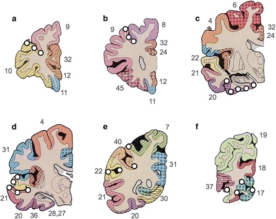
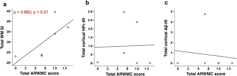
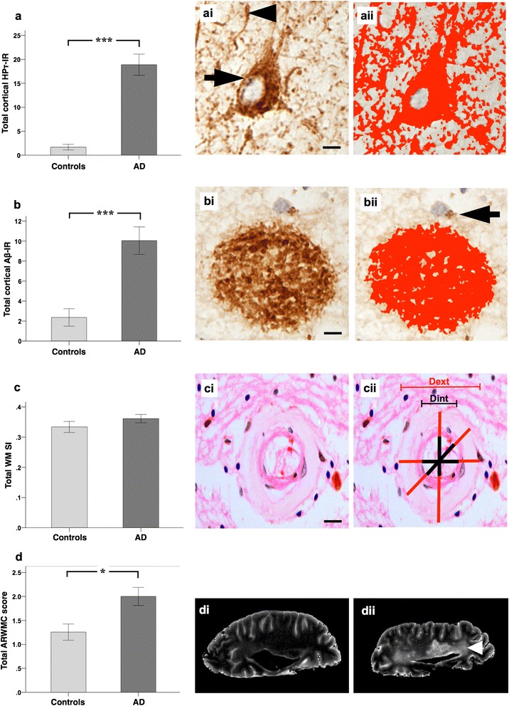
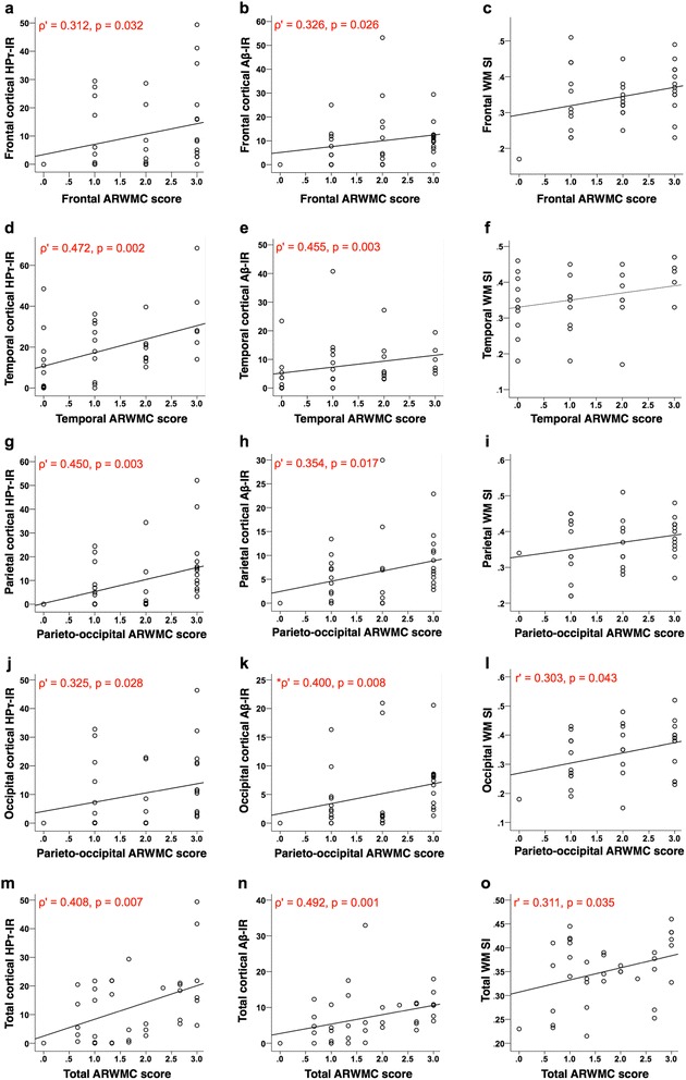
36 human post-mortem right fixed cerebral hemispheres (mean age 84.4 ± 7.7 years; male: 16, female: 20) containing varying amounts of AD-pathology (AD: 23, controls: 13) underwent T2- weighted MRI with WMH assessed according to the age related white matter change scale (ARWMC). After dissection, using tissue samples from the frontal, temporal, parietal and occipital regions from the right hemisphere, we quantitatively assessed cortical HPτ and Aβ pathology burden by measuring the percentage area covered by AT8 immunoreactivity (HPτ-IR) and 4G8 immunoreactivity (Aβ-IR), and assessed the severity of WM SVD by calculating the sclerotic index (SI) of WM arteries/arterioles. HPτ-IR, Aβ-IR, and SI were compared with ARWMC scores. HPτ-IR, Aβ-IR and WM ARWMC scores were all significantly higher in AD cases compared to controls, while SI values were similar between groups. ARWMC scores correlated with HPτ-IR, Aβ-IR and SI in various regions, however, linear regression revealed that only HPτ-IR was a significant independent predictor of ARWMC scores.
Here we have shown that increasing cortical HPτ burden independently predicted the severity of WMH indicating its potentially important role in the pathogenesis of WM damage. Moreover, our findings suggest that in AD patients the presence of WMH may indicate cortical AD-associated pathology rather than SVD. Further studies are warranted to elucidate the pathological processes that lead to WM damage and to clarify if WMH may serve as a general biomarker for cortical AD-associated pathology.
脑白质病变(WML),在 T2 加权 MRI 上表现为脑白质高信号(WMH),包含脑白质(WM)的结构损伤和完整性丧失,通常被认为与小血管疾病(SVD)有关。然而,有人认为 WM 损伤也可能是皮质阿尔茨海默病(AD)病理即过度磷酸化的 tau(HPτ)和淀粉样β(Aβ)继发的轴突退行性丢失的结果。在这里,我们研究了 HPτ、Aβ 和 SVD 对 WMH 严重程度的影响。
36 个人死后右侧固定大脑半球(平均年龄 84.4±7.7 岁;男性 16 例,女性 20 例)包含不同数量的 AD 病理(AD:23 例,对照组:13 例)接受 T2 加权 MRI,根据年龄相关性白质改变量表(ARWMC)评估 WMH。在解剖后,使用来自右侧半球额、颞、顶和枕叶的组织样本,我们通过测量 AT8 免疫反应性(HPτ-IR)和 4G8 免疫反应性(Aβ-IR)的覆盖面积百分比来定量评估皮质 HPτ 和 Aβ 病理负担,并通过计算 WM 动脉/小动脉的硬化指数(SI)来评估 WM SVD 的严重程度。将 HPτ-IR、Aβ-IR 和 WM SVD 与 ARWMC 评分进行比较。与对照组相比,AD 病例的 HPτ-IR、Aβ-IR 和 WM ARWMC 评分均显著升高,而两组之间的 SI 值相似。ARWMC 评分与各区域的 HPτ-IR、Aβ-IR 和 SI 均相关,但线性回归显示仅 HPτ-IR 是 ARWMC 评分的显著独立预测因子。
在这里,我们表明皮质 HPτ 负荷的增加独立预测了 WMH 的严重程度,表明其在 WM 损伤发病机制中具有潜在的重要作用。此外,我们的发现表明,在 AD 患者中,WMH 的存在可能表明与皮质 AD 相关的病理,而不是 SVD。需要进一步的研究来阐明导致 WM 损伤的病理过程,并阐明 WMH 是否可以作为皮质 AD 相关病理的一般生物标志物。