Tefera Endale, Bermudez-Cañete Ramon, van Doorn Carin
Cardiology Unit, Department of Pediatrics and Child Heath, School of Medicine, Addis Ababa University, Corner of Zambia and T. Abanefso road, P.O.Box 1768, Addis Ababa, Ethiopia.
Department of Pediatric Cardiology, Ramon y Cajal University Hospital, Madrid, Spain.
BMC Res Notes. 2015 Sep 30;8:511. doi: 10.1186/s13104-015-1467-3.
Inadvertent ligation of the left pulmonary artery during attempted surgical closure of a Patent Ductus Arteriosus has long been recognized as one of the less common complications of this procedure. Surgical reconstruction of the left pulmonary artery was then often attempted but was difficult or impossible in some of the patients with hypoplasia of the left pulmonary artery and the left lung.
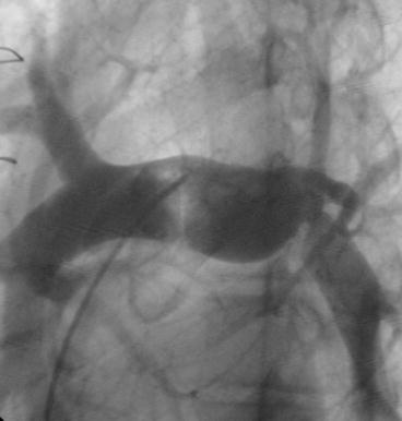
A 10-year-old girl presented with marked exercise intolerance and palpitations and was diagnosed to have large PDA. She had feeding difficulty, diaphoresis, failure to gain weight, recurrent chest infections during infancy and early childhood. Physical examination revealed an underweight child with wide pulse pressure and bounding peripheral pulses. She had active precordium with accentuated P2 and machinery murmur in the left 2nd intercostal space and mid diastolic rumble at the mitral area. Echocardiography showed a 12 mm patent arterial duct. She was taken for an intended surgical ligation of the duct but a control echocardiogram on the 3rd postoperative day revealed that the left pulmonary artery, instead of the duct, was ligated. Surgical reconstruction of the left pulmonary artery was undertaken 3 years later, however, this was complicated by post reconstruction left pulmonary artery stenosis. Successful percutaneous stenting of the left pulmonary artery was performed 18 months after the surgical reconstruction.
The incidence of inadvertent LPA ligation may be underestimated where PDA ligation is done by less experienced surgeons and postoperative echocardiography is not routinely performed. Late correction of inadvertent LPA ligation is an important surgical challenge, especially if the duct is still patent. Percutaneous stenting as a primary option may carry significant risk, as the ligated pulmonary artery is fragile. In our case, a good result was achieved with surgical repair followed by percutaneous stenting.
在尝试手术闭合动脉导管未闭时意外结扎左肺动脉一直被认为是该手术较少见的并发症之一。当时常常尝试对左肺动脉进行手术重建,但对于一些左肺动脉和左肺发育不全的患者来说,手术重建困难甚至无法进行。
一名10岁女孩因明显运动不耐受和心悸就诊,被诊断为大型动脉导管未闭。她在婴儿期和幼儿期有喂养困难、多汗、体重不增、反复肺部感染。体格检查发现患儿体重不足,脉压增宽,外周脉搏搏动增强。心前区活跃,P2亢进,左第2肋间有连续性机器样杂音,二尖瓣区有舒张中期隆隆样杂音。超声心动图显示动脉导管未闭直径为12毫米。她接受了动脉导管预期结扎手术,但术后第3天的对照超声心动图显示结扎的是左肺动脉而非动脉导管。3年后对左肺动脉进行了手术重建,然而,重建后出现了左肺动脉狭窄并发症。手术重建18个月后成功进行了经皮左肺动脉支架置入术。
在由经验不足的外科医生进行动脉导管结扎且未常规进行术后超声心动图检查的情况下,意外结扎左肺动脉的发生率可能被低估。意外结扎左肺动脉的晚期矫正手术是一项重要的外科挑战,尤其是在动脉导管仍未闭的情况下。经皮支架置入作为首选方案可能存在重大风险,因为结扎后的肺动脉很脆弱。在我们的病例中,先进行手术修复然后经皮支架置入取得了良好效果。