Tachibana Yasuhiko, Obata Takayuki, Tsuchiya Hiroki, Omatsu Tokuhiko, Kishimoto Riwa, Kawaguchi Hiroshi, Nishikori Akira, Kamagata Koji, Hori Masaaki, Aoki Shigeki, Tsuji Hiroshi, Inoue Tomio
Research Center for Charged Particle Therapy, National Institute of Radiological Sciences, 4-9-1 Anagawa, Inage-ku, Chiba, 263-8555, Japan.
Department of Radiology, Yokohama City University Graduate School of Medicine, 3-9 Fukuura, Kanazawa-ku, Yokohama, 236-0004, Japan.
Eur Radiol. 2016 Aug;26(8):2559-66. doi: 10.1007/s00330-015-4038-z. Epub 2015 Oct 7.
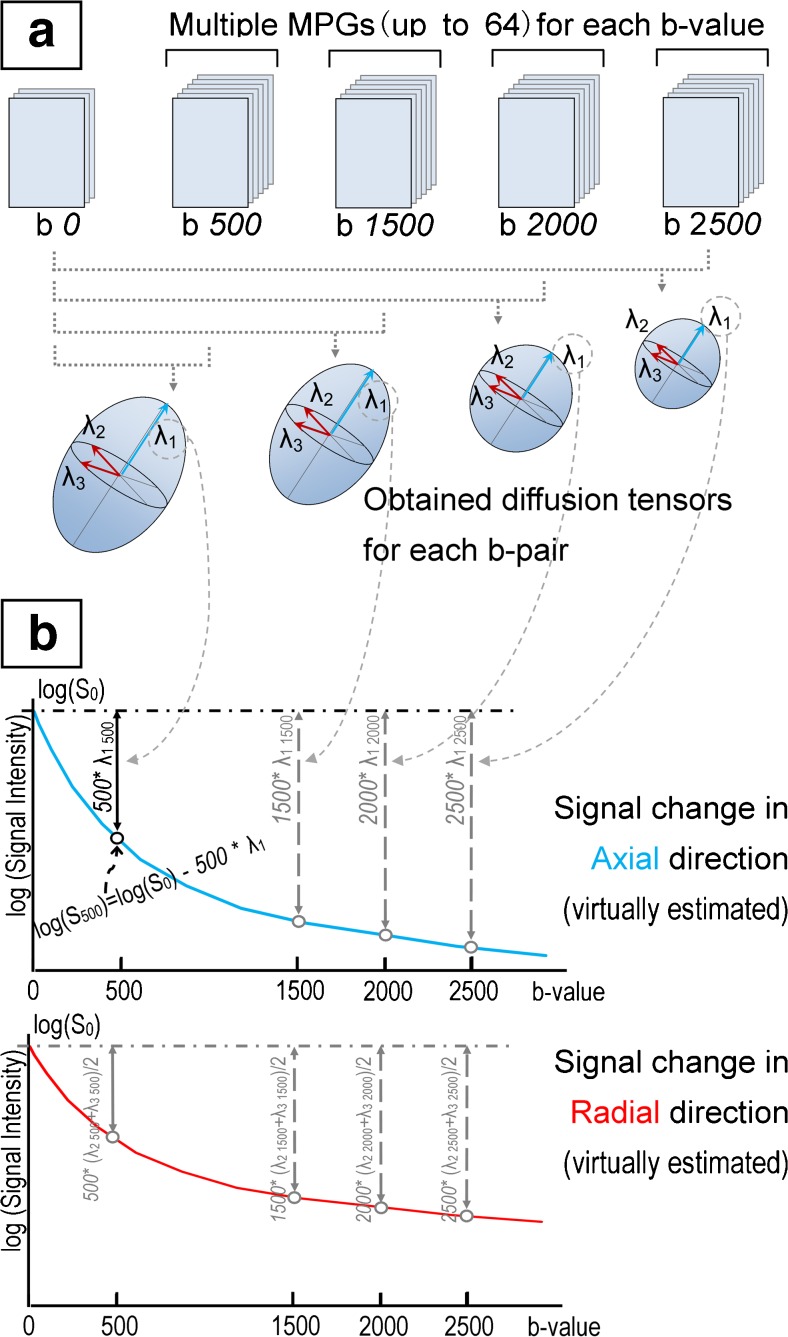
A new method that can estimate diffusional kurtosis image (DKI), estimated DKI (eDKI), parallel and perpendicular to neuronal fibres from greatly limited image data was designed to enable quick and practical assessment of DKI in clinics. The purpose of this study was to discuss the potential of this method for clinical use.
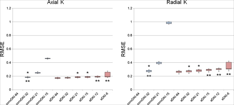
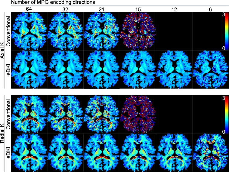
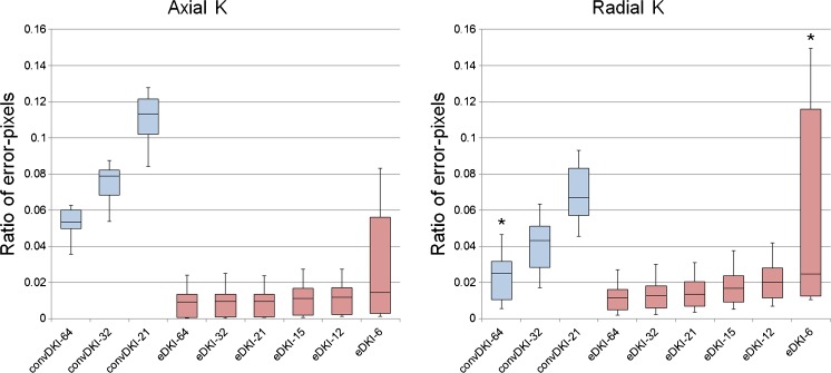
Fourteen healthy volunteers were examined with a 3-Tesla MRI. The diffusion-weighting parameters included five different b-values (0, 500, 1,500, 2,000 and 2,500 s/mm(2)) with 64 different encoding directions for each of the b-values. K values were calculated by both conventional DKI (convDKI) and eDKI from these complete data, and also from the data that the encoding directions were abstracted to 32, 21, 15, 12 and 6. Error-pixel ratio and the root mean square error (RMSE) compared with the standard were compared between the methods (Wilcoxon signed-rank test: P < 0.05 was considered significant).
Error-pixel ratio was smaller in eDKI than in convDKI and the difference was significant. In addition, RMSE was significantly smaller in eDKI than in convDKI, or otherwise the differences were not significant when they were obtained from the same data set.
eDKI might be useful for assessing DKI in clinical settings.
• A method to practically estimate axial/radial DKI from limited data was developed. • The high robustness of the proposed method can greatly improve map images. • The accuracy of the proposed method was high. • Axial/radial K maps can be calculated from limited diffusion-encoding directions. • The proposed method might be useful for assessing DKI in clinical settings.
设计一种新方法,能够从非常有限的图像数据中估计扩散峰度图像(DKI)、估计DKI(eDKI),并平行和垂直于神经纤维,以实现临床中DKI的快速和实用评估。本研究的目的是探讨该方法在临床应用中的潜力。
对14名健康志愿者进行3特斯拉磁共振成像检查。扩散加权参数包括五个不同的b值(0、500、1500、2000和2500 s/mm²),每个b值有64个不同的编码方向。通过传统DKI(convDKI)和eDKI从这些完整数据中计算K值,也从编码方向抽象为32、21、15、12和6的数据中计算K值。比较各方法与标准相比的误差像素比和均方根误差(RMSE)(Wilcoxon符号秩检验:P < 0.05被认为具有显著性)。
eDKI的误差像素比小于convDKI,差异具有显著性。此外,eDKI的RMSE显著小于convDKI,或者当从同一数据集获得时差异不显著。
eDKI可能有助于在临床环境中评估DKI。
• 开发了一种从有限数据中实际估计轴向/径向DKI的方法。• 所提出方法的高稳健性可大大改善图谱图像。• 所提出方法的准确性高。• 可从有限的扩散编码方向计算轴向/径向K图。• 所提出的方法可能有助于在临床环境中评估DKI。