Pacioni Simone, D'Alessandris Quintino Giorgio, Giannetti Stefano, Morgante Liliana, De Pascalis Ivana, Coccè Valentina, Bonomi Arianna, Pascucci Luisa, Alessandri Giulio, Pessina Augusto, Falchetti Maria Laura, Pallini Roberto
Institute of Neurosurgery, Università Cattolica del Sacro Cuore, Largo Agostino Gemelli 8, 00168, Rome, Italy.
CNR-Institute of Cell Biology and Neurobiology (IBCN), via del Fosso di Fiorano 64, 00143, Rome, Italy.
Stem Cell Res Ther. 2015 Oct 6;6:194. doi: 10.1186/s13287-015-0185-z.
The goal of cancer chemotherapy is targeting tumor cells and/or tumor-associated microvessels with the lowest systemic toxicity. Mesenchymal stromal cells (MSCs) are promising vehicles for selective drug delivery due to their peculiar ability to home to pathological tissues. We previously showed that MSCs are able to uptake and subsequently to release the chemotherapeutic compound Paclitaxel (PTX) and to impair the growth of subcutaneous glioblastoma multiforme (GBM) xenografts. Here we used an orthotopic GBM model 1) to assess whether PTX-loaded MSCs (PTX-MSCs) retain a tropism towards the tumor cells in the brain context, and 2) to characterize the cytotoxic damage induced by MSCs-driven PTX release in the tumor microenvironment.
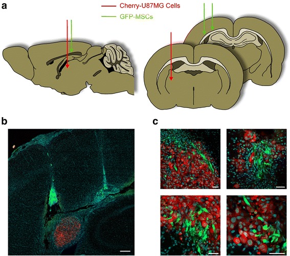
U87MG GBM cells were fluorescently labeled with the mCherry protein and grafted onto the brain of immunosuppressed rats. In adjacent brain regions, we injected green fluorescent protein-expressing murine MSCs, either loaded with PTX or unloaded. After 1 week survival, the xenografted brain was assessed by confocal microscopy for PTX-induced cell damage.
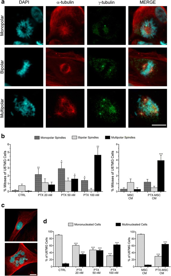
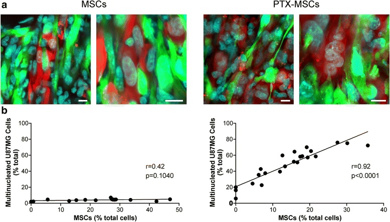
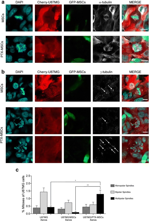
Overall, MSCs showed remarkable tropism towards the tumor. In rats grafted with PTX-MSCs, the nuclei of U87MG cells showed changes that are typically induced by PTX, including multi-spindle mitoses, centrosome number alterations, and nuclear fragmentation. Multi-spindle mitoses resulted in multinucleated cells that were significantly higher in tumors co-grafted with PTX-MSCs than in controls. Nuclear changes did not occur in astrocytes and neurons surrounding the tumor.
MSCs appear particularly suited for anti-neoplastic drug delivery in the brain since PTX-specific damage of GBM cells can be achieved avoiding side effects to the normal tissue.
癌症化疗的目标是以最低的全身毒性靶向肿瘤细胞和/或肿瘤相关微血管。间充质基质细胞(MSC)因其具有归巢至病理组织的独特能力,是选择性药物递送的理想载体。我们之前表明,MSC能够摄取并随后释放化疗化合物紫杉醇(PTX),并抑制皮下多形性胶质母细胞瘤(GBM)异种移植物的生长。在此,我们使用原位GBM模型:1)评估负载PTX的MSC(PTX-MSC)在脑环境中是否仍对肿瘤细胞具有趋向性;2)表征由MSC驱动的PTX在肿瘤微环境中释放所诱导的细胞毒性损伤。
用mCherry蛋白对U87MG GBM细胞进行荧光标记,并将其移植到免疫抑制大鼠的脑中。在相邻的脑区,我们注射了表达绿色荧光蛋白的小鼠MSC,其中一部分负载PTX,一部分未负载。存活1周后,通过共聚焦显微镜评估移植了异种移植物的脑内PTX诱导的细胞损伤。
总体而言,MSC对肿瘤表现出显著的趋向性。在移植了PTX-MSC的大鼠中,U87MG细胞的细胞核显示出通常由PTX诱导的变化,包括多极有丝分裂、中心体数量改变和核碎裂。多极有丝分裂导致多核细胞的出现,在与PTX-MSC共同移植的肿瘤中,多核细胞显著多于对照组。肿瘤周围的星形胶质细胞和神经元未出现核变化。
MSC似乎特别适合在脑内进行抗肿瘤药物递送,因为可以实现GBM细胞的PTX特异性损伤,同时避免对正常组织产生副作用。