Jungen Christiane, Zeus Tobias, Balzer Jan, Eickholt Christian, Petersen Margot, Kehmeier Eva, Veulemans Verena, Kelm Malte, Willems Stephan, Meyer Christian
Department of Cardiology-Electrophysiology, cNEP, cardiac Neuro- and Electrophysiology research group, University Heart Center, University Hospital Hamburg-Eppendorf, Hamburg, Germany, DZHK (German Center for Cardiovascular Research), partner site Hamburg/Kiel/Luebeck, Hamburg, Germany; Department of Cardiology, Pulmonology and Vascular Medicine, Medical Faculty, University Hospital Duesseldorf, Duesseldorf, Germany.
Department of Cardiology, Pulmonology and Vascular Medicine, Medical Faculty, University Hospital Duesseldorf, Duesseldorf, Germany.
PLoS One. 2015 Oct 14;10(10):e0140386. doi: 10.1371/journal.pone.0140386. eCollection 2015.
To investigate whether percutaneous left atrial appendage (LAA) closure guided by automated real-time integration of 2D-/3D-transesophageal echocardiography (TEE) and fluoroscopy imaging results in decreased radiation exposure.
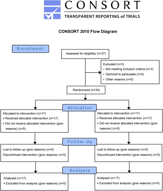
In this open-label single-center study LAA closure (AmplatzerTM Cardiac Plug) was performed in 34 consecutive patients (8 women; 73.1±8.5 years) with (n = 17, EN+) or without (n = 17, EN-) integrated echocardiography/fluoroscopy imaging guidance (EchoNavigator® [EN]; Philips Healthcare). There were no significant differences in baseline characteristics between both groups. Successful LAA closure was documented in all patients. Radiation dose was reduced in the EN+ group about 52% (EN+: 48.5±30.7 vs. EN-: 93.9±64.4 Gy/cm2; p = 0.01). Corresponding to the radiation dose fluoroscopy time was reduced (EN+: 16.7±7 vs. EN-: 24.0±11.4 min; p = 0.035). These advantages were not at the cost of increased procedure time (89.6±28.8 vs. 90.1±30.2 min; p = 0.96) or periprocedural complications. Contrast media amount was comparable between both groups (172.3±92.7 vs. 197.5±127.8 ml; p = 0.53). During short-term follow-up of at least 3 months (mean: 8.1±5.9 months) no device-related events occurred.
Automated real-time integration of echocardiography and fluoroscopy can be incorporated into procedural work-flow of percutaneous left atrial appendage closure without prolonging procedure time. This approach results in a relevant reduction of radiation exposure.
ClinicalTrials.gov NCT01262508.
研究二维/三维经食管超声心动图(TEE)与荧光透视成像的自动实时整合引导下的经皮左心耳(LAA)封堵术是否能减少辐射暴露。
在这项开放标签的单中心研究中,连续34例患者(8名女性;73.1±8.5岁)接受了LAA封堵术(AmplatzerTM心脏封堵器),其中17例(EN+组)采用了整合超声心动图/荧光透视成像引导(EchoNavigator® [EN];飞利浦医疗保健公司),另外17例(EN-组)未采用。两组患者的基线特征无显著差异。所有患者的LAA封堵均获成功。EN+组的辐射剂量降低了约52%(EN+组:48.5±30.7 vs. EN-组:93.9±64.4 Gy/cm2;p = 0.01)。与辐射剂量相应,荧光透视时间也缩短了(EN+组:16.7±7 vs. EN-组:24.0±11.4分钟;p = 0.035)。这些优势并未以增加手术时间(89.6±28.8 vs. 90.1±30.2分钟;p = 0.96)或围手术期并发症为代价。两组的造影剂用量相当(172.3±92.7 vs. 197.5±127.8毫升;p = 0.53)。在至少3个月(平均:8.1±5.9个月)的短期随访中,未发生与器械相关的事件。
超声心动图与荧光透视的自动实时整合可纳入经皮左心耳封堵术的手术流程,且不延长手术时间。这种方法可显著减少辐射暴露。
ClinicalTrials.gov NCT01262508。