Cho Chan-Ho, Lee Sang-Bumm
Wolhang Public Health Center, # 1151, Anpo-ri, Wolhang-myeon, Seongju-gun, Gyeongsang-bukdo, 719-851, South Korea.
Department of Ophthalmology, Yeungnam University College of Medicine, #170, Hyunchung-ro, Nam-gu, Daegu, 705-717, South Korea.
BMC Ophthalmol. 2015 Oct 24;15:140. doi: 10.1186/s12886-015-0130-z.
Scleromalacia, in the form of scleral thinning, melting, and necrosis, is a potentially serious complication of pterygium excision. This study introduces a new biodegradable material, Ologen™ collagen matrix (OCM), to repair scleral thinning as an alternative to preserved scleral tissue, and evaluates the long-term outcomes of OCM for ocular surface reconstruction surgery.
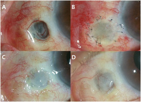
Two cases of possibly mitomycin C (MMC)-associated marked scleral thinning after pterygium excision with 0.02 % topical MMC for 2-weeks were included in this study. An OCM graft at the scleral thinning area and conjunctival autograft (CAU) were performed on both patients. The scleral defect size was measured and its margin was marked with a biopsy punch. The margin of the scleral thinning area was trimmed by Vannas scissors and the OCM was cut using a circular-shape biopsy punch of the same size. The OCM was sutured with a recipient scleral wall using 10-0 nylon interrupted sutures. Free CAU was harvested from the superonasal bulbar conjunctiva with a punch biopsy 1-mm larger in diameter than that of the OCM. The previously sutured OCM bed was covered with CAU and the graft was secured with 10-0 nylon interrupted sutures. Both patients were examined periodically for over two years by assessing graft thickness and surface vascularization using a slit lamp biomicroscope. Reepithelialization of the ocular surface was observed within three to six days after surgery. Ocular discomfort and inflammation ceased in both patients as the ocular surface quickly stabilized. The entire graft site remained intact and provided a good healthy ocular surface with fluorescein stain negative intact epithelium and good vascularization of grafted conjunctiva. Epithelial defects and scleral thinning did not recur in either patient over the two year follow-up period.
For treatment of a possibly MMC-associated scleral necrosis following the surgical excision of the pterygium, an OCM graft with CAU is highly recommended for good clinical outcomes and low recurrence rates. With the clinical results of this study, the new biodegradable Ologen™ collagen matrix qualifies as an alternative treatment to scleral tissue for ocular surface reconstruction.
巩膜软化表现为巩膜变薄、溶解和坏死,是翼状胬肉切除术后一种潜在的严重并发症。本研究引入一种新型可生物降解材料Ologen™胶原基质(OCM),用于修复巩膜变薄,作为保存巩膜组织的替代方法,并评估OCM用于眼表重建手术的长期效果。
本研究纳入2例在翼状胬肉切除术后使用0.02%局部丝裂霉素C(MMC)治疗2周后可能与MMC相关的显著巩膜变薄病例。对2例患者均在巩膜变薄区域进行OCM移植及结膜自体移植(CAU)。测量巩膜缺损大小,并用活检打孔器标记其边缘。用Vannas剪刀修剪巩膜变薄区域的边缘,并用相同大小的圆形活检打孔器切割OCM。用10-0尼龙间断缝线将OCM与受体巩膜壁缝合。从鼻上球结膜用直径比OCM大1mm的活检打孔器获取游离CAU。将先前缝合的OCM床覆盖CAU,并用10-0尼龙间断缝线固定移植片。使用裂隙灯生物显微镜评估移植片厚度和表面血管化情况,对2例患者进行了两年多的定期检查。术后3至6天观察到眼表重新上皮化。随着眼表迅速稳定,2例患者的眼部不适和炎症均消失。整个移植部位保持完整,荧光素染色显示上皮完整阴性,移植结膜血管化良好,提供了健康的眼表。在两年的随访期内,2例患者均未出现上皮缺损和巩膜变薄复发。
对于翼状胬肉手术切除后可能与MMC相关的巩膜坏死的治疗,强烈推荐使用OCM移植联合CAU,以获得良好的临床效果和低复发率。基于本研究的临床结果,新型可生物降解的Ologen™胶原基质有资格作为眼表重建中巩膜组织的替代治疗方法。