Chong Shau Poh, Merkle Conrad W, Cooke Dylan F, Zhang Tingwei, Radhakrishnan Harsha, Krubitzer Leah, Srinivasan Vivek J
Opt Lett. 2015 Nov 1;40(21):4911-4. doi: 10.1364/OL.40.004911.
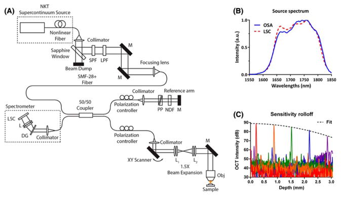
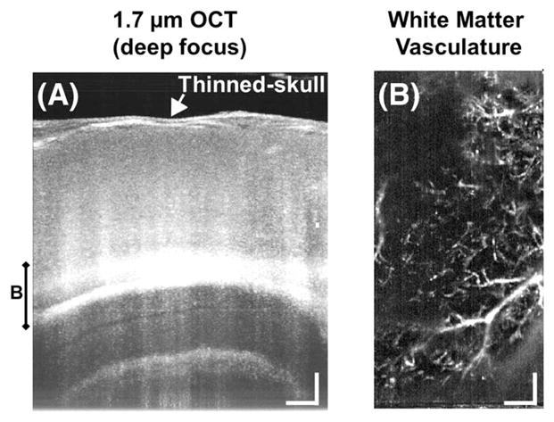
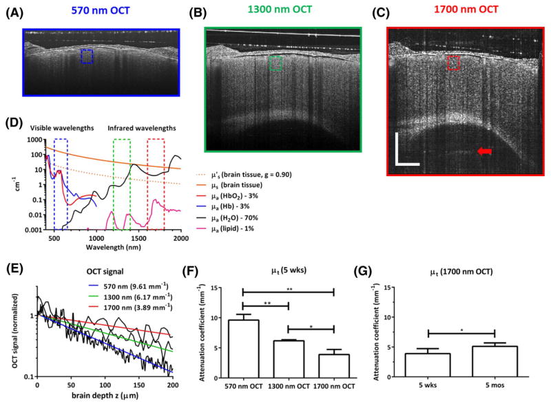
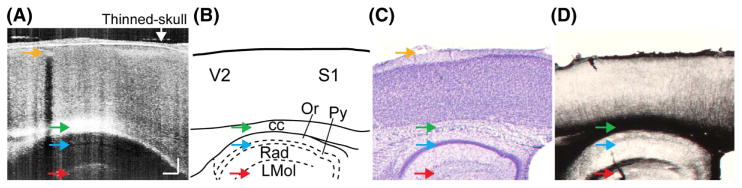
A spectral/Fourier domain optical coherence tomography (OCT) intravital microscope using a supercontinuum light source at 1.7 μm was developed to study subcortical structures noninvasively in the living mouse brain. The benefits of 1.7 μm for deep tissue brain imaging are demonstrated by quantitatively comparing OCT signal attenuation characteristics of cortical tissue across visible and near-infrared wavelengths. Imaging of hippocampal tissue architecture and white matter microvasculature are demonstrated in vivo through thinned-skull, glass coverslip-reinforced cranial windows in mice. Applications of this novel platform include monitoring disease progression and pathophysiology in rodent models of Alzheimer's disease and subcortical dementias, including vascular dementia.
开发了一种使用1.7μm超连续光源的光谱/傅里叶域光学相干断层扫描(OCT)活体显微镜,用于在活体小鼠大脑中无创地研究皮层下结构。通过定量比较可见和近红外波长下皮质组织的OCT信号衰减特性,证明了1.7μm对深部脑组织成像的益处。通过小鼠薄颅骨、玻璃盖玻片加固的颅窗,在体内展示了海马组织结构和白质微血管系统的成像。这个新平台的应用包括监测阿尔茨海默病和皮层下痴呆(包括血管性痴呆)啮齿动物模型中的疾病进展和病理生理学。