Gassert Robert B, Pearson William G
Department of Cellular Biology and Anatomy, Medical College of Georgia at Georgia Regents University, 1120 15th Street, Augusta, GA 30912, USA.
Magn Reson Imaging. 2016 Feb;34(2):204-8. doi: 10.1016/j.mri.2015.10.029. Epub 2015 Oct 31.
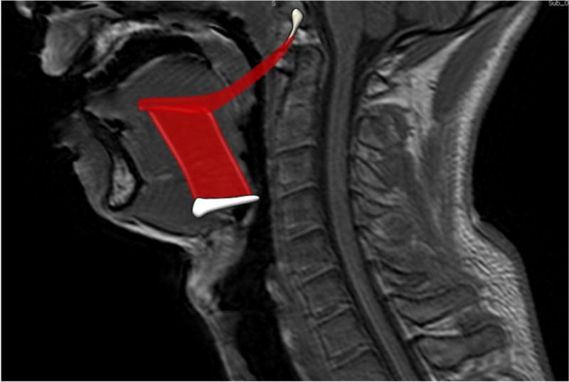
Tongue base retraction during swallowing is critical to bolus propulsion in normal physiological swallowing. A better understanding of the hyoglossus and styloglossus, muscles thought to be key to tongue base retraction, will improve the quality of physical rehabilitation in dysphagic patients in addition to preventing iatrogenic damage to structures critical to deglutition. This study utilized muscle functional MRI in healthy adult human subjects in order to determine if the hyoglossus and styloglossus are active during swallowing.
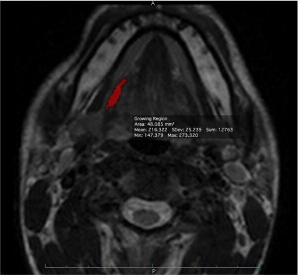
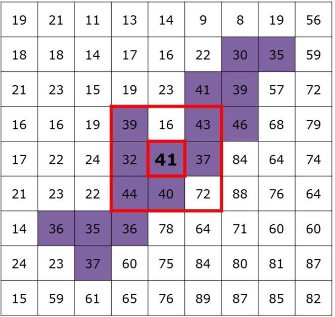
Data were collected for 11 subjects with mfMRI before and after swallowing, and after performing the Mendelsohn maneuver. Whole muscle relaxation time profiles (T2 signal in milliseconds) were calculated from weighted averages of multiple dual echo MRI slices, allowing for comparison of physiological response for the muscles in each test condition. Changes in effect size (Cohen's d) of whole muscle T2 profiles were used to establish whether or not the hyoglossus and styloglossus are utilized during swallowing and during the Mendelsohn maneuver.
Post-swallowing effect size changes (where a d value of >0.20 indicates significant activity) for the T2 signal profiles of the hyoglossus and styloglossus were found to be d=1.19 and 0.22, respectively. The hyoglossus showed an effect size change of d=0.26 for the Mendelsohn maneuver.
Muscle functional MRI indicates a physiological response of the hyoglossus and styloglossus during swallowing, and the hyoglossus during the Mendelsohn maneuver.
吞咽过程中舌根后缩对于正常生理吞咽时食团推进至关重要。更好地了解被认为是舌根后缩关键的舌骨舌肌和茎突舌肌,除了可防止对吞咽关键结构造成医源性损伤外,还将提高吞咽困难患者的物理康复质量。本研究对健康成年受试者使用肌肉功能磁共振成像,以确定舌骨舌肌和茎突舌肌在吞咽过程中是否活跃。
收集了11名受试者在吞咽前后以及进行门德尔松手法操作后的mfMRI数据。通过多个双回波MRI切片的加权平均值计算全肌弛豫时间曲线(以毫秒为单位的T2信号),以便比较每种测试条件下肌肉的生理反应。使用全肌T2曲线的效应大小变化(科恩d值)来确定舌骨舌肌和茎突舌肌在吞咽过程中和门德尔松手法操作期间是否被使用。
发现吞咽后舌骨舌肌和茎突舌肌T2信号曲线的效应大小变化(d值>0.20表示有显著活动)分别为d = 1.19和0.22。舌骨舌肌在门德尔松手法操作时的效应大小变化为d = 0.26。
肌肉功能磁共振成像表明吞咽过程中舌骨舌肌和茎突舌肌有生理反应,门德尔松手法操作时舌骨舌肌有生理反应。