Department of Ophthalmology, Laboratory for Experimental Immunology of the Eye, University of Cologne, 50931, Cologne, Germany.
Centre for Experimental Medicine, School of Medicine, Dentistry and Biomedical Sciences, Queen's University Belfast, Belfast, BT12 6BA, UK.
J Neuroinflammation. 2015 Nov 2;12:201. doi: 10.1186/s12974-015-0422-5.
Reactive microglia are commonly seen in retinal degenerative diseases, and neurotoxic microglia responses can contribute to photoreceptor cell death. We and others have previously shown that translocator protein (18 kDa) (TSPO) is highly induced in retinal degenerations and that the selective TSPO ligand XBD173 (AC-5216, emapunil) exerts strong anti-inflammatory effects on microglia in vitro and ex vivo. Here, we investigated whether targeting TSPO with XBD173 has immuno-modulatory and neuroprotective functions in two mouse models of acute retinal degeneration using bright white light exposure.
BALB/cJ and Cx3cr1(GFP/+) mice received intraperitoneal injections of 10 mg/kg XBD173 or vehicle for five consecutive days, starting 1 day prior to exposure to either 15,000 lux white light for 1 h or 50,000 lux focal light for 10 min, respectively. The effects of XBD173 treatment on microglia and Müller cell reactivity were analyzed by immuno-stainings of retinal sections and flat mounts, fluorescence-activated cell sorting (FACS) analysis, and mRNA expression of microglia markers using quantitative real-time PCR (qRT-PCR). Optical coherence tomography (OCT), terminal deoxynucleotidyl transferase dUTP nick end labeling (TUNEL) stainings, and morphometric analyses were used to quantify the extent of retinal degeneration and photoreceptor apoptosis.
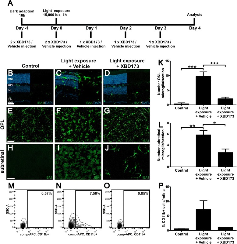
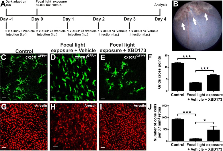
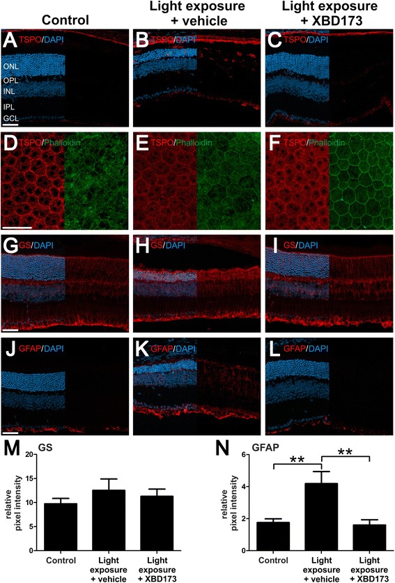
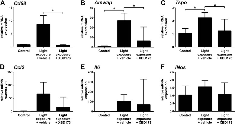
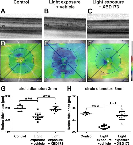
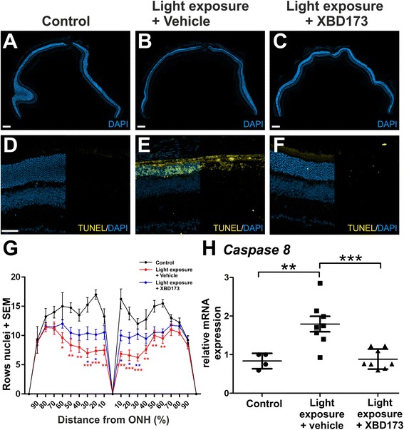
Four days after the mice were challenged with bright white light, a large number of amoeboid-shaped alerted microglia appeared in the degenerating outer retina, which was nearly completely prevented by treatment with XBD173. This treatment also down-regulated the expression of TSPO protein in microglia but did not change the TSPO levels in the retinal pigment epithelium (RPE). RT-PCR analysis showed that the microglia/macrophage markers Cd68 and activated microglia/macrophage whey acidic protein (Amwap) as well as the pro-inflammatory genes Ccl2 and Il6 were reduced after XBD173 treatment. Light-induced degeneration of the outer retina was nearly fully blocked by XBD173 treatment. We further confirmed these findings in an independent mouse model of focal light damage. Retinas of animals receiving XBD173 therapy displayed significantly more ramified non-reactive microglia and more viable arrestin-positive cone photoreceptors than vehicle controls.
Targeting TSPO with XBD173 effectively counter-regulates microgliosis and ameliorates light-induced retinal damage, highlighting a new pharmacological concept for the treatment of retinal degenerations.
反应性小胶质细胞常见于视网膜退行性疾病中,神经毒性小胶质细胞反应可能导致光感受器细胞死亡。我们和其他人之前已经表明,转位蛋白(18 kDa)(TSPO)在视网膜变性中高度诱导,并且选择性 TSPO 配体 XBD173(AC-5216,emapunil)在体外和离体对小胶质细胞具有强烈的抗炎作用。在这里,我们研究了使用明亮的白光照射,用 XBD173 靶向 TSPO 是否在两种急性视网膜变性的小鼠模型中具有免疫调节和神经保护功能。
BALB/cJ 和 Cx3cr1(GFP/+) 小鼠在暴露于 15,000 lux 白光 1 小时或 50,000 lux 焦点光 10 分钟之前,每天连续腹膜内注射 10 mg/kg XBD173 或载体,连续 5 天。通过视网膜切片和平面贴片中的免疫染色、荧光激活细胞分选(FACS)分析以及使用定量实时 PCR(qRT-PCR)检测小胶质细胞标志物的 mRNA 表达,分析 XBD173 治疗对小胶质细胞和 Müller 细胞反应性的影响。光相干断层扫描(OCT)、末端脱氧核苷酸转移酶 dUTP 缺口末端标记(TUNEL)染色和形态计量分析用于量化视网膜变性和光感受器细胞凋亡的程度。
在小鼠受到明亮的白光刺激四天后,大量变形的醒着的小胶质细胞出现在变性的外视网膜中,用 XBD173 治疗几乎完全阻止了这种情况。这种治疗还下调了小胶质细胞中 TSPO 蛋白的表达,但没有改变视网膜色素上皮(RPE)中的 TSPO 水平。RT-PCR 分析显示,小胶质细胞/巨噬细胞标志物 Cd68 和激活的小胶质细胞/巨噬细胞乳酸性蛋白(Amwap)以及促炎基因 Ccl2 和 Il6 在 XBD173 治疗后减少。用 XBD173 治疗几乎完全阻断了外视网膜的光诱导变性。我们在独立的焦点光损伤小鼠模型中进一步证实了这些发现。接受 XBD173 治疗的动物的视网膜显示出更多的分支非反应性小胶质细胞和更多的存活的 arrestin 阳性视锥细胞,而载体对照则较少。
用 XBD173 靶向 TSPO 可有效调节小胶质细胞增生并改善光诱导的视网膜损伤,突出了治疗视网膜变性的新药理学概念。