Bagheri Fatemeh, Safarian Shahrokh, Eslaminejad Mohamadreza Baghaban, Sheibani Nader
a Department of Cell and Molecular Biology, School of Biology, College of Science, University of Tehran, Tehran, Iran.
b Department of Stem Cells and Developmental Biology, Cell Sciences Research Center, Royan Institute for Stem Cell Biology and Technology, ACECR, Tehran, Iran.
Biochem Cell Biol. 2015 Dec;93(6):604-10. doi: 10.1139/bcb-2015-0007. Epub 2015 Sep 15.
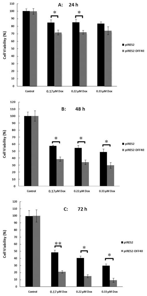
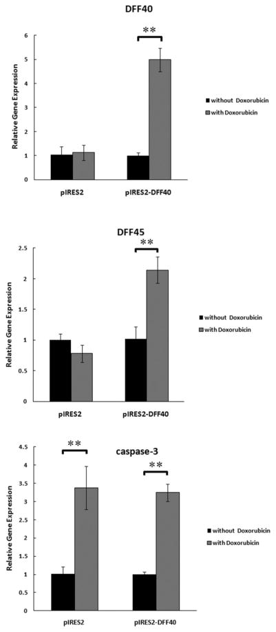
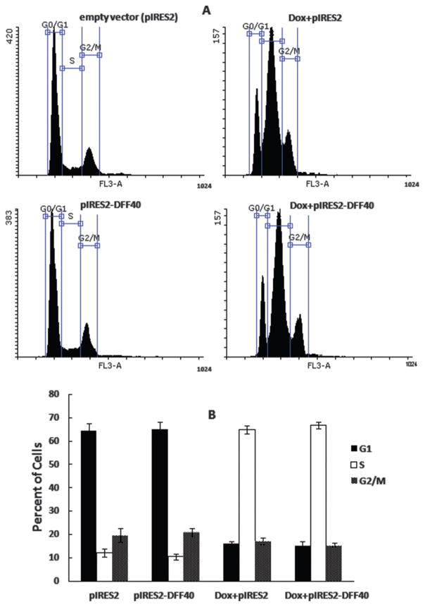
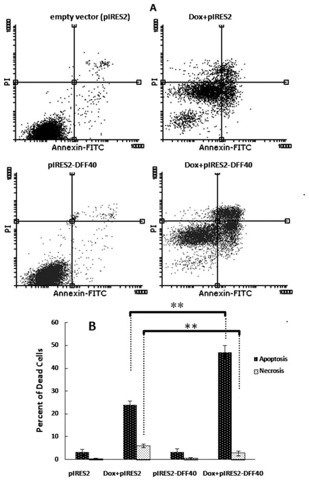
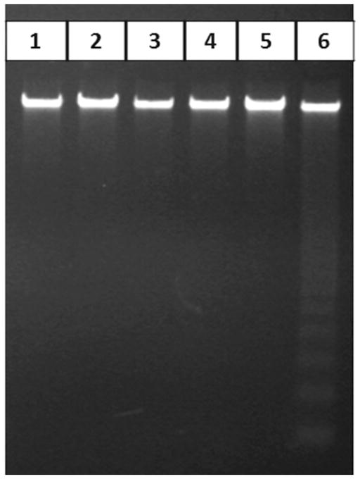
There are a number of reports demonstrating a relationship between the alterations in DFF40 expression and development of some cancers. Here, increased DFF40 expression in T-47D cells in the presence of doxorubicin was envisaged for therapeutic usage. The T-47D cells were transfected with an eukaryotic expression vector encoding the DFF40 cDNA. Following incubation with doxorubicin, propidium iodide (PI) staining was used for cell cycle distribution analysis. The rates of apoptosis were determined by annexin V/PI staining. Apoptosis was also evaluated using the DNA laddering analysis. The viability of DFF40-transfected cells incubated with doxorubicin was significantly decreased compared with control cells. However, there were no substantial changes in the cell cycle distribution of pIRES2-DFF40 cells incubated with doxorubicin compared to control cells. The expression of DFF40, without doxorubicin incubation, had also no significant effect on the cell cycle distribution. There was no DNA laddering in cells transfected with the empty pIRES2 vector when incubated with doxorubicin. In contrast, DNA laddering was observed in DFF40 transfected cells in the presence of doxorubicin after 48 h. Also, the expression of DFF40 and DFF45 was increased in DFF40 transfected cells in the presence of doxorubicin enhancing cell death. Collectively our results indicated that co-treatment of DFF40-transfected cells with doxorubicin can enhance the killing of these tumor cells via apoptosis. Thus, modulation of DFF40 level may be a beneficial strategy for treatment of chemo-resistant cancers.
有许多报告表明DFF40表达的改变与某些癌症的发生发展之间存在关联。在此,设想在阿霉素存在的情况下,T-47D细胞中DFF40表达的增加可用于治疗用途。用编码DFF40 cDNA的真核表达载体转染T-47D细胞。在与阿霉素孵育后,使用碘化丙啶(PI)染色进行细胞周期分布分析。通过膜联蛋白V/PI染色确定凋亡率。还使用DNA梯状分析评估凋亡情况。与对照细胞相比,用阿霉素孵育的DFF40转染细胞的活力显著降低。然而,与对照细胞相比,用阿霉素孵育的pIRES2-DFF40细胞的细胞周期分布没有实质性变化。在不进行阿霉素孵育的情况下,DFF40的表达对细胞周期分布也没有显著影响。用空的pIRES2载体转染的细胞在与阿霉素孵育时没有DNA梯状条带。相反,在阿霉素存在的情况下,48小时后在DFF40转染细胞中观察到DNA梯状条带。此外,在阿霉素存在的情况下,DFF40转染细胞中DFF40和DFF45的表达增加,增强了细胞死亡。总体而言,我们的结果表明,DFF40转染细胞与阿霉素联合处理可通过凋亡增强对这些肿瘤细胞的杀伤作用。因此,调节DFF40水平可能是治疗化疗耐药癌症的有益策略。