Investigation performed at the Department of Orthopedics and Traumatology, Faculty of Medicine, University of São Paulo, São Paulo, Brazil ; Department of Orthopaedics and Traumatology, Institute of Orthopedics and Traumatology-Hospital and Clinics, Faculty of Medicine, University of São Paulo (IOT-HCFMUSP), São Paulo, Brazil.
Department of Orthopaedics and Traumatology, Institute of Orthopedics and Traumatology-Hospital and Clinics, Faculty of Medicine, University of São Paulo (IOT-HCFMUSP), São Paulo, Brazil.
Orthop J Sports Med. 2013 Dec 9;1(7):2325967113513546. doi: 10.1177/2325967113513546. eCollection 2013 Dec.
Reconstruction of the anterior cruciate ligament (ACL) is one of the most common procedures in orthopaedic surgery. However, even with advances in surgical techniques and implants, some patients still have residual anterolateral rotatory laxity after reconstruction. A thorough study of the anatomy of the anterolateral region of the knee is needed.
To study the anterolateral region and determine the measurements and points of attachments of the anterolateral ligament (ALL).
Descriptive laboratory study.
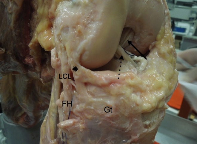
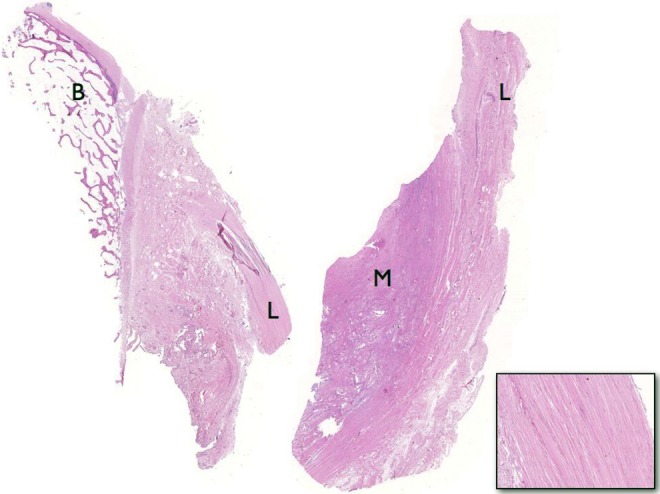
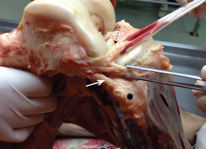
Dissections of the anterolateral structures of the knee were performed in 20 human cadavers. After isolating the ALL, its length, thickness, width, and points of attachments were determined. The femoral attachment of the ALL was based on the anterior-posterior and proximal-distal distances from the attachment of the lateral collateral ligament (LCL). The tibial attachment point was based on the distance from the Gerdy tubercle to the fibular head and the distance from the lateral tibial plateau. The ligaments from the first 10 dissections were sent for histological analysis.
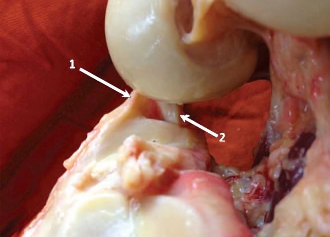
The ALL was found in all 20 knees. The femoral attachment of the ALL at the lateral epicondyle averaged 3.5 mm distal and 2.2 mm anterior to the attachment of the LCL. Two distal attachments were observed: one inserts into the lateral meniscus, the other between the Gerdy tubercle and the fibular head, approximately 4.4 mm distal to the tibial articular cartilage. The mean measurements for the ligament were 37.3 mm (length), 7.4 mm (width), and 2.7 mm (thickness). The histological analysis of the ligaments revealed dense connective tissue.
The ALL is consistently present in the anterolateral region of the knee. Its attachment to the femur is anterior and distal to the attachment of the LCL. Moving distally, it bifurcates at close to half of its length. The ALL features 2 distal attachments, one at the lateral meniscus and the other between the Gerdy tubercle and the fibular head.
The ALL may be important in maintaining normal rotatory limits of knee motion; ALL rupture could be responsible for rotatory laxity after isolated intra-articular reconstruction of the ACL.
重建前交叉韧带(ACL)是矫形外科中最常见的手术之一。然而,即使手术技术和植入物有所进步,一些患者在重建后仍存在残余前外侧旋转松弛。需要对膝关节前外侧区域进行深入解剖研究。
研究前外侧区域并确定前外侧韧带(ALL)的测量和附着点。
描述性实验室研究。
对 20 具人体尸体的膝关节前外侧结构进行解剖。在分离 ALL 后,确定其长度、厚度、宽度和附着点。ALL 的股骨附着点基于从外侧副韧带(LCL)附着点的前后和远近距离。胫骨附着点基于从 Gerdy 结节到腓骨头的距离和从胫骨外侧平台的距离。前 10 次解剖的韧带被送去进行组织学分析。
在所有 20 个膝关节中均发现了 ALL。ALL 在外侧髁的股骨附着点平均距离 LCL 附着点 3.5 毫米远和 2.2 毫米前。观察到两个远端附着点:一个插入外侧半月板,另一个位于 Gerdy 结节和腓骨头之间,距胫骨关节软骨约 4.4 毫米远。韧带的平均测量值为 37.3 毫米(长度)、7.4 毫米(宽度)和 2.7 毫米(厚度)。韧带的组织学分析显示为致密结缔组织。
ALL 始终存在于膝关节的前外侧区域。其在股骨上的附着点位于 LCL 附着点的前方和远侧。向远端移动时,它在接近一半的长度处分叉。ALL 有两个远端附着点,一个在外侧半月板上,另一个在 Gerdy 结节和腓骨头之间。
ALL 可能对维持膝关节正常旋转运动范围很重要;ALL 断裂可能是 ACL 关节内重建后旋转松弛的原因。