Roy Paromita, Chakraborty Sayantani, Das Sudip, Roy Alok
Department of ENT, National Medical College, NRS Medical College, Kolkata, West Bengal, India.
Department of Dermatology, Venereology and Leprosy, NRS Medical College, Kolkata, West Bengal, India.
Indian J Dermatol. 2015 Sep-Oct;60(5):497-9. doi: 10.4103/0019-5154.164374.
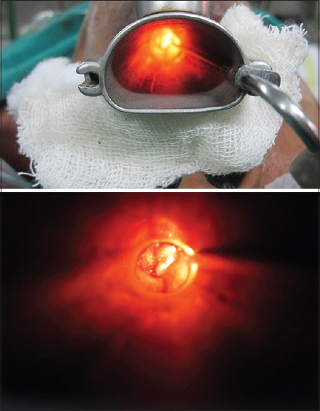
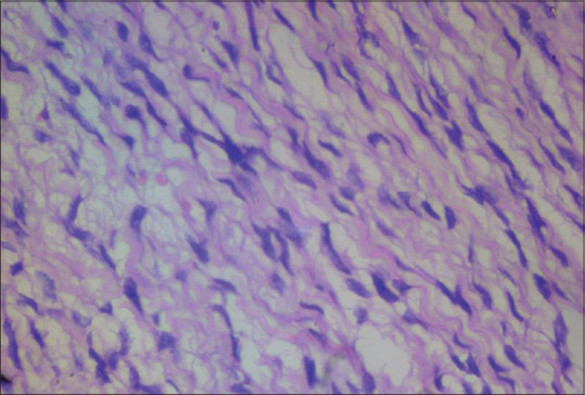
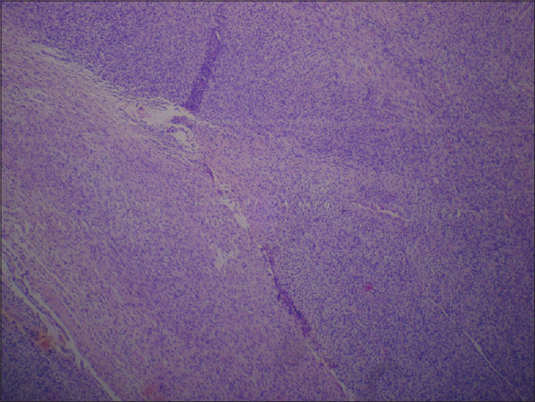
A 75-year-old man presented with a slowly growing mass at the right side of the base of the tongue for 4 months. The mass was painless initially but had become very painful during preceding 4 weeks. On examination a 3 cm diameter, oval swelling was observed at the right side of the base of the tongue. It was firm in consistency, slightly tender, non-ulcerative, and with irregular surface. A deep incisional biopsy was taken from mass under general anesthesia. Histopathology report identified the mass consistent with neurofibroma. It featured typical pallisading arrangement of fascicles of spindle-shaped cells and there was no evidence of malignancy. An absence of Verocay body and thick hyalinized vessels ruled out Schwannoma. No similar lesions were found in any other part of the patient's body. He exhibited no skin pigmentation, no hearing deficit, and no evidence suggestive of any systemic disorders that might have been attributable to the tongue base neurofibroma. His family history was also negative. Thus, a diagnosis of isolated neurofibroma of the tongue was established. The patient was advised excision of the mass but he refused and lost in follow up.
一名75岁男性因舌根部右侧有一缓慢生长的肿物4个月前来就诊。该肿物起初无痛,但在之前4周变得非常疼痛。检查发现舌根部右侧有一个直径3厘米的椭圆形肿胀。质地坚硬,稍有压痛,无溃疡,表面不规则。在全身麻醉下从肿物处取了深部切口活检。组织病理学报告显示该肿物符合神经纤维瘤。其特征为梭形细胞束典型的栅栏状排列,且无恶性证据。无Verocay小体和增厚的玻璃样变血管排除了神经鞘瘤。在患者身体的任何其他部位均未发现类似病变。他没有皮肤色素沉着,没有听力缺陷,也没有任何可能归因于舌根部神经纤维瘤的全身性疾病的迹象。他的家族史也为阴性。因此,确诊为孤立性舌神经纤维瘤。建议患者切除肿物,但他拒绝了,随后失访。