Nyante Sarah J, Sherman Mark E, Pfeiffer Ruth M, Berrington de Gonzalez Amy, Brinton Louise A, Bowles Erin J Aiello, Hoover Robert N, Glass Andrew, Gierach Gretchen L
Division of Cancer Epidemiology and Genetics, National Cancer Institute, Rockville, Maryland.
Division of Cancer Epidemiology and Genetics, National Cancer Institute, Rockville, Maryland. Division of Cancer Prevention, National Cancer Institute, Rockville, Maryland.
Cancer Epidemiol Biomarkers Prev. 2016 Jan;25(1):212-6. doi: 10.1158/1055-9965.EPI-15-0412. Epub 2015 Nov 6.
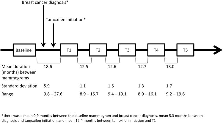
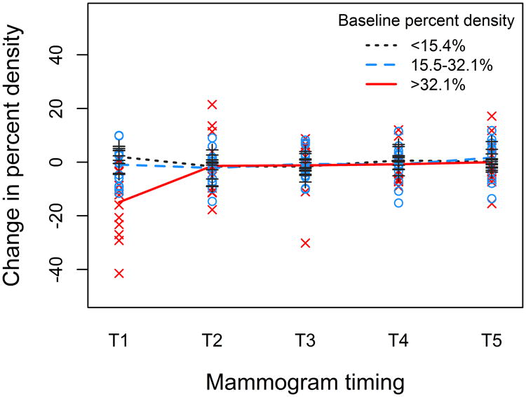
Tamoxifen-associated mammographic density (MD) reductions are linked to improved breast cancer survival. We evaluated MD at six time points to determine the timing of greatest reduction following tamoxifen initiation. We sampled 40 Kaiser Permanente Northwest estrogen receptor (ER)-positive breast cancer patients from a prior study of MD change, according to tamoxifen use duration and age at diagnosis: <4 years tamoxifen and ≤50 years (N = 6) or >50 years (N = 10) old; ≥4 years tamoxifen and ≤50 years (N = 13) or >50 years (N = 11) old. A single reader evaluated percent MD in the contralateral breast on baseline (pre-diagnosis) and five approximately yearly post-diagnostic (T1 to T5) mammograms. Mean MD change was calculated. Interactions with age (≤50 and >50 years), tamoxifen duration (<4 and ≥4 years), and baseline MD (tertiles) were tested in linear regression models. Overall, the largest MD decline occurred by T1 (mean 4.5%) with little additional decline by T5. Declines differed by tertile of baseline MD (Pinteraction < 0.01). In the highest tertile, the largest reduction occurred by T1 (mean 14.9%), with an additional reduction of 3.6% by T5. Changes were smaller in the middle and lowest baseline MD tertiles, with cumulative reductions of 3.0% and 0.4% from baseline to T5, respectively. There were no differences by age (Pinteraction = 0.36) or tamoxifen duration (Pinteraction = 0.42). Among ER-positive patients treated with tamoxifen and surviving ≥5 years, most of the MD reduction occurred within approximately 12 months of tamoxifen initiation, suggesting that MD measurement at a single time point following tamoxifen initiation can identify patients with substantial density declines.
他莫昔芬相关的乳腺钼靶密度(MD)降低与乳腺癌生存率提高有关。我们在六个时间点评估了MD,以确定开始使用他莫昔芬后MD最大程度降低的时间。我们根据他莫昔芬使用时长和诊断时的年龄,从先前一项关于MD变化的研究中抽取了40名凯撒医疗西北分部雌激素受体(ER)阳性乳腺癌患者:使用他莫昔芬<4年且≤50岁(N = 6)或>50岁(N = 10);使用他莫昔芬≥4年且≤50岁(N = 13)或>50岁(N = 11)。由一名阅片者评估对侧乳房在基线(诊断前)以及诊断后的五张大致每年一次的乳房X光片(T1至T5)上的MD百分比。计算平均MD变化。在线性回归模型中检验了年龄(≤50岁和>50岁)、他莫昔芬使用时长(<4年和≥4年)以及基线MD(三分位数)之间的相互作用。总体而言,MD最大降幅出现在T1时(平均4.5%),到T5时几乎没有额外降幅。降幅因基线MD三分位数而异(P相互作用<0.01)。在最高三分位数中,最大降幅出现在T1时(平均14.9%),到T5时又额外降低了3.6%。基线MD处于中间和最低三分位数时变化较小,从基线到T5的累积降幅分别为3.0%和0.4%。年龄(P相互作用 = 0.36)或他莫昔芬使用时长(P相互作用 = 0.42)方面无差异。在接受他莫昔芬治疗且存活≥5年的ER阳性患者中,大部分MD降低发生在开始使用他莫昔芬后的大约12个月内,这表明在开始使用他莫昔芬后的单个时间点进行MD测量可以识别出密度有显著下降的患者。