Zaccagnini Germana, Palmisano Anna, Canu Tamara, Maimone Biagina, Lo Russo Francesco M, Ambrogi Federico, Gaetano Carlo, De Cobelli Francesco, Del Maschio Alessandro, Esposito Antonio, Martelli Fabio
Molecular Cardiology Laboratory, IRCCS-Policlinico San Donato, San Donato Milanese, Milan, Italy.
Preclinical Imaging Facility, Experimental Imaging Center, San Raffaele Scientific Institute, Milan, Italy.
PLoS One. 2015 Nov 10;10(11):e0142111. doi: 10.1371/journal.pone.0142111. eCollection 2015.
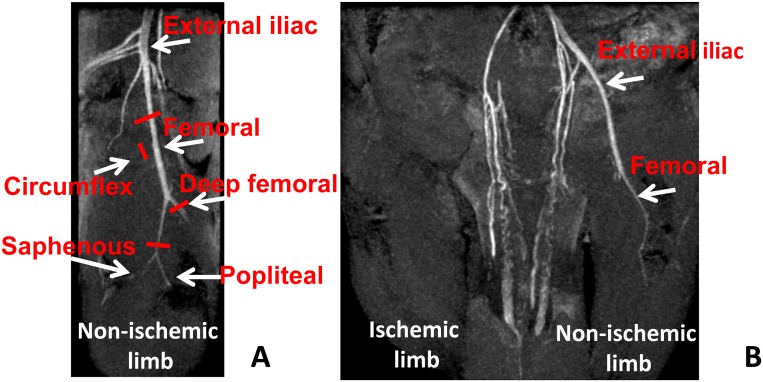
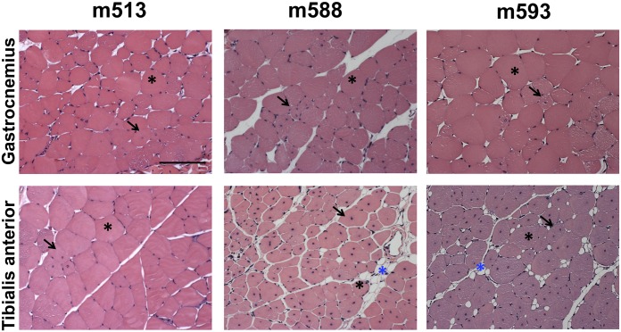
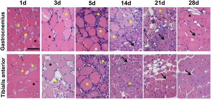
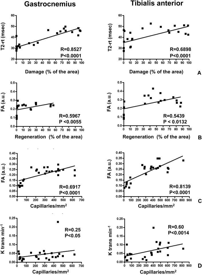
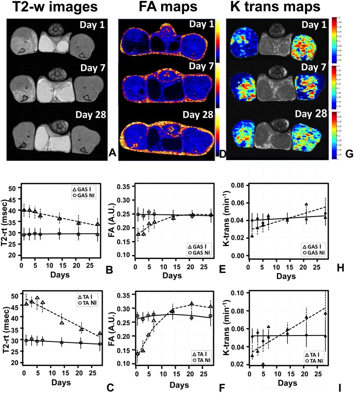
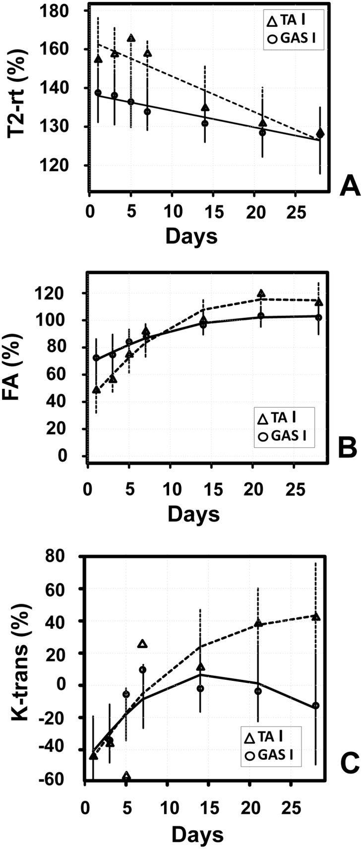
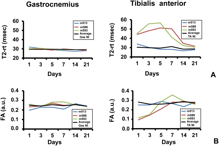
Magnetic resonance imaging (MRI) provides non-invasive, repetitive measures in the same individual, allowing the study of a physio-pathological event over time. In this study, we tested the performance of 7 Tesla multi-parametric MRI to monitor the dynamic changes of mouse skeletal muscle injury and regeneration upon acute ischemia induced by femoral artery dissection. T2-mapping (T2 relaxation time), diffusion-tensor imaging (Fractional Anisotropy) and perfusion by Dynamic Contrast-Enhanced MRI (K-trans) were measured and imaging results were correlated with histological morphometric analysis in both Gastrocnemius and Tibialis anterior muscles. We found that tissue damage positively correlated with T2-relaxation time, while myofiber regeneration and capillary density positively correlated with Fractional Anisotropy. Interestingly, K-trans positively correlated with capillary density. Accordingly, repeated MRI measurements between day 1 and day 28 after surgery in ischemic muscles showed that: 1) T2-relaxation time rapidly increased upon ischemia and then gradually declined, returning almost to basal level in the last phases of the regeneration process; 2) Fractional Anisotropy dropped upon ischemic damage induction and then recovered along with muscle regeneration and neoangiogenesis; 3) K-trans reached a minimum upon ischemia, then progressively recovered. Overall, Gastrocnemius and Tibialis anterior muscles displayed similar patterns of MRI parameters dynamic, with more marked responses and less variability in Tibialis anterior. We conclude that MRI provides quantitative information about both tissue damage after ischemia and the subsequent vascular and muscle regeneration, accounting for the differences between subjects and, within the same individual, between different muscles.
磁共振成像(MRI)可在同一个体中提供非侵入性的重复测量,从而能够对生理病理事件进行长期研究。在本研究中,我们测试了7特斯拉多参数MRI监测股动脉剥离诱导急性缺血后小鼠骨骼肌损伤和再生动态变化的性能。测量了T2映射(T2弛豫时间)、扩散张量成像(分数各向异性)以及动态对比增强MRI灌注(Ktrans),并将成像结果与腓肠肌和胫骨前肌的组织形态计量学分析进行了关联。我们发现组织损伤与T2弛豫时间呈正相关,而肌纤维再生和毛细血管密度与分数各向异性呈正相关。有趣的是,Ktrans与毛细血管密度呈正相关。因此,对缺血肌肉术后第1天至第28天进行的重复MRI测量结果显示:1)T2弛豫时间在缺血后迅速增加,随后逐渐下降,在再生过程的最后阶段几乎恢复到基础水平;2)分数各向异性在缺血损伤诱导后下降,然后随着肌肉再生和新生血管形成而恢复;3)Ktrans在缺血时达到最小值,然后逐渐恢复。总体而言,腓肠肌和胫骨前肌的MRI参数动态变化模式相似,胫骨前肌的反应更明显且变异性更小。我们得出结论,MRI提供了有关缺血后组织损伤以及随后血管和肌肉再生的定量信息,解释了不同个体之间以及同一个体内不同肌肉之间的差异。