• 文献检索
  • 文档翻译
  • 深度研究
  • 学术资讯
  • Suppr Zotero 插件Zotero 插件
  • 邀请有礼
  • 套餐&价格
  • 历史记录
应用&插件
Suppr Zotero 插件Zotero 插件浏览器插件Mac 客户端Windows 客户端微信小程序
定价
高级版会员购买积分包购买API积分包
服务
文献检索文档翻译深度研究API 文档MCP 服务
关于我们
关于 Suppr公司介绍联系我们用户协议隐私条款
关注我们

Suppr 超能文献

核心技术专利:CN118964589B侵权必究
粤ICP备2023148730 号-1Suppr @ 2026

文献检索

告别复杂PubMed语法,用中文像聊天一样搜索,搜遍4000万医学文献。AI智能推荐,让科研检索更轻松。

立即免费搜索

文件翻译

保留排版,准确专业,支持PDF/Word/PPT等文件格式,支持 12+语言互译。

免费翻译文档

深度研究

AI帮你快速写综述,25分钟生成高质量综述,智能提取关键信息,辅助科研写作。

立即免费体验

体外培养类型会影响恶性疟原虫的细胞黏附、凸起结构、蛋白质定位和转录组图谱。

Type of in vitro cultivation influences cytoadhesion, knob structure, protein localization and transcriptome profile of Plasmodium falciparum.

作者信息

Tilly Ann-Kathrin, Thiede Jenny, Metwally Nahla, Lubiana Pedro, Bachmann Anna, Roeder Thomas, Rockliffe Nichola, Lorenzen Stephan, Tannich Egbert, Gutsmann Thomas, Bruchhaus Iris

机构信息

Bernhard Nocht Institute for Tropical Medicine, Bernhard-Nocht-Str. 74, 20359 Hamburg, Germany.

Zoological Institute, Molecular Physiology, Christian-Albrechts University Kiel, Olshausenstraße 40, 24098 Kiel, Germany.

出版信息

Sci Rep. 2015 Nov 16;5:16766. doi: 10.1038/srep16766.

DOI:10.1038/srep16766
PMID:26568166
原文链接:https://pmc.ncbi.nlm.nih.gov/articles/PMC4645185/
Abstract

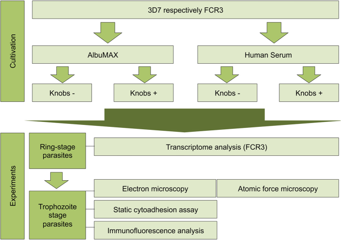
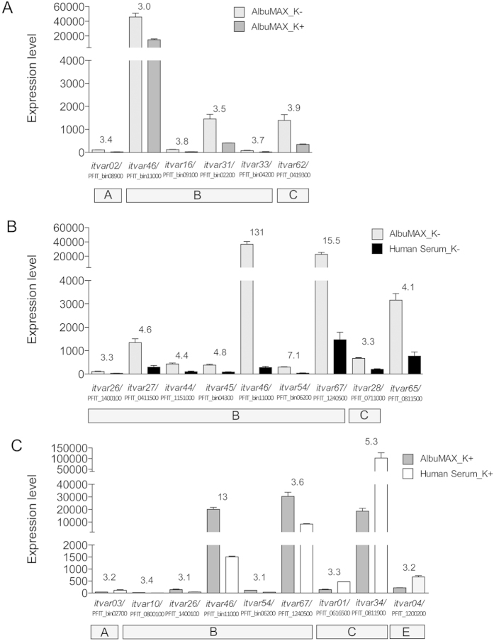
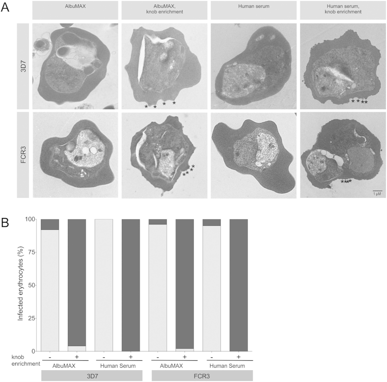
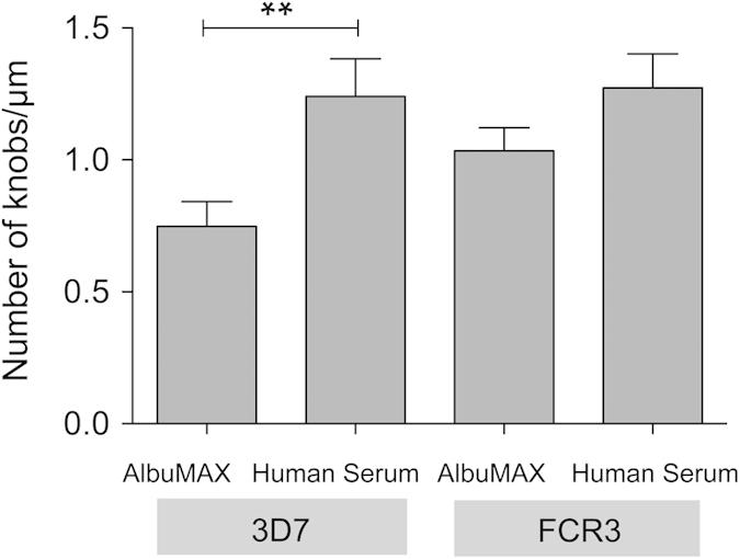
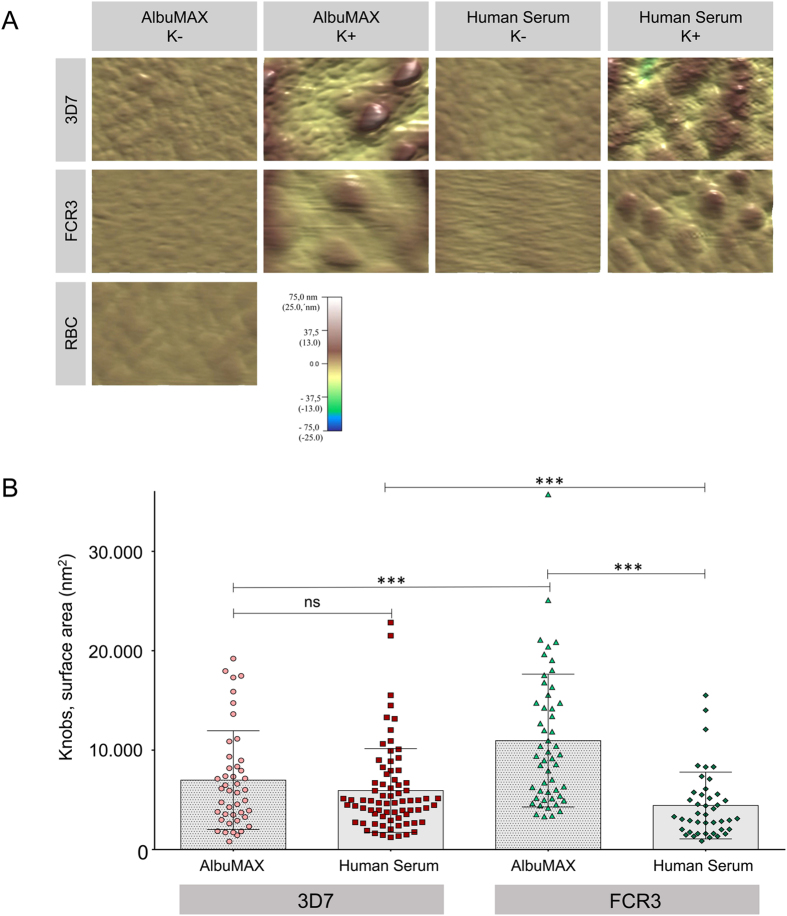
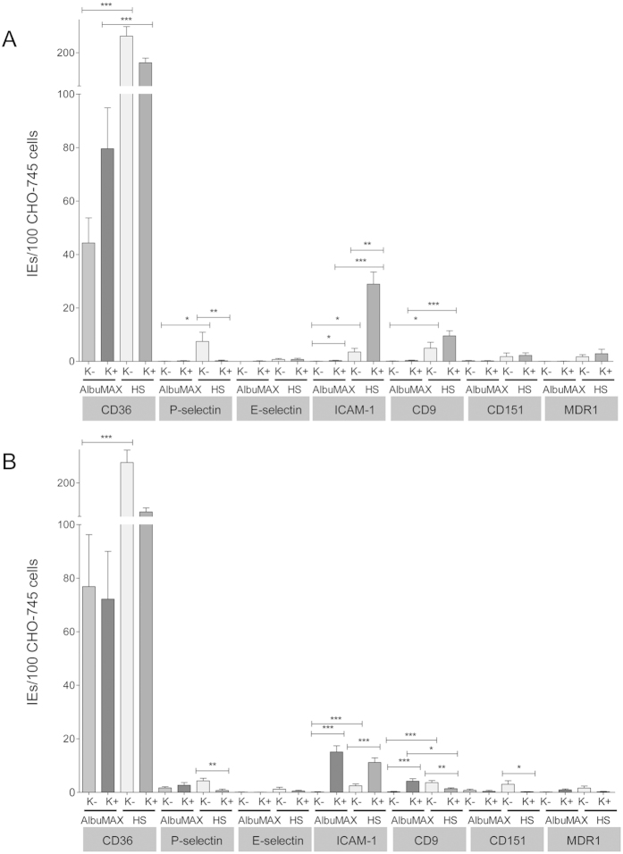
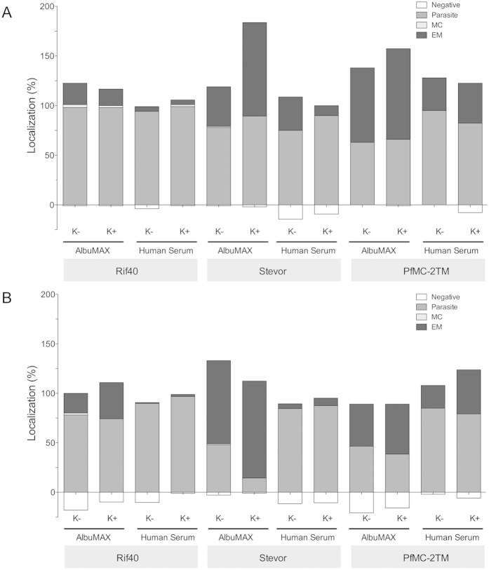
In vitro cultivation of Plasmodium falciparum is critical for studying the biology of this parasite. However, it is likely that different in vitro cultivation conditions influence various aspects of the parasite's life cycle. In the present study two P. falciparum isolates were cultivated using the two most common methods, in which AlbuMAX or human serum as additives are used, and the results were compared. The type of cultivation influenced the knob structure of P. falciparum-infected erythrocytes (IEs). IEs cultivated with AlbuMAX had fewer knobs than those cultivated with human serum. Furthermore, knob size varied between isolates and is also depended on the culture medium. In addition, there was a greater reduction in the cytoadhesion of IEs to various endothelial receptors in the presence of AlbuMAX than in the presence of human serum. Surprisingly, cytoadhesion did not correlate with the presence or absence of knobs. Greater numbers of the variant surface antigen families RIFIN, STEVOR, and PfMC-2TM were found at the IE membrane when cultivated in the presence of AlbuMAX. Moreover, the type of cultivation had a marked influence on the transcriptome profile. Compared with cultivation with human serum, cultivation with AlbuMAX increased the expression of approximately 500-870 genes.

摘要

恶性疟原虫的体外培养对于研究该寄生虫的生物学特性至关重要。然而,不同的体外培养条件可能会影响寄生虫生命周期的各个方面。在本研究中,使用两种最常用的方法培养了两株恶性疟原虫分离株,这两种方法分别使用白蛋白(AlbuMAX)或人血清作为添加剂,并对结果进行了比较。培养类型影响了恶性疟原虫感染红细胞(IEs)的凸起结构。用AlbuMAX培养的IEs的凸起比用人血清培养的少。此外,不同分离株的凸起大小不同,并且还取决于培养基。此外,与用人血清培养相比,在AlbuMAX存在的情况下,IEs与各种内皮受体的细胞黏附性降低得更多。令人惊讶的是,细胞黏附性与凸起的有无无关。在AlbuMAX存在的情况下培养时,在IE膜上发现了更多数量的可变表面抗原家族RIFIN、STEVOR和PfMC-2TM。此外,培养类型对转录组图谱有显著影响。与用人血清培养相比,用AlbuMAX培养使大约500 - 870个基因的表达增加。

https://cdn.ncbi.nlm.nih.gov/pmc/blobs/401d/4645185/8a57c5d92e76/srep16766-f7.jpg
https://cdn.ncbi.nlm.nih.gov/pmc/blobs/401d/4645185/4347dc8b280a/srep16766-f1.jpg
https://cdn.ncbi.nlm.nih.gov/pmc/blobs/401d/4645185/cd8916e16343/srep16766-f2.jpg
https://cdn.ncbi.nlm.nih.gov/pmc/blobs/401d/4645185/3bc9e24f4115/srep16766-f3.jpg
https://cdn.ncbi.nlm.nih.gov/pmc/blobs/401d/4645185/44956c353147/srep16766-f4.jpg
https://cdn.ncbi.nlm.nih.gov/pmc/blobs/401d/4645185/355425309e02/srep16766-f5.jpg
https://cdn.ncbi.nlm.nih.gov/pmc/blobs/401d/4645185/2fc04e1b0ded/srep16766-f6.jpg
https://cdn.ncbi.nlm.nih.gov/pmc/blobs/401d/4645185/8a57c5d92e76/srep16766-f7.jpg

相似文献

1
Type of in vitro cultivation influences cytoadhesion, knob structure, protein localization and transcriptome profile of Plasmodium falciparum.体外培养类型会影响恶性疟原虫的细胞黏附、凸起结构、蛋白质定位和转录组图谱。
Sci Rep. 2015 Nov 16;5:16766. doi: 10.1038/srep16766.
2
A comparative study of the localization and membrane topology of members of the RIFIN, STEVOR and PfMC-2TM protein families in Plasmodium falciparum-infected erythrocytes.恶性疟原虫感染红细胞中RIFIN、STEVOR和PfMC-2TM蛋白家族成员的定位及膜拓扑结构的比较研究
Malar J. 2015 Jul 16;14:274. doi: 10.1186/s12936-015-0784-2.
3
Temporal expression and localization patterns of variant surface antigens in clinical Plasmodium falciparum isolates during erythrocyte schizogony.红细胞裂殖期临床疟原虫分离株变异表面抗原的时间表达和定位模式。
PLoS One. 2012;7(11):e49540. doi: 10.1371/journal.pone.0049540. Epub 2012 Nov 15.
4
Expression switching in the stevor and Pfmc-2TM superfamilies in Plasmodium falciparum.恶性疟原虫中stevor和Pfmc-2TM超家族的表达转换
Mol Microbiol. 2007 Jun;64(6):1621-34. doi: 10.1111/j.1365-2958.2007.05767.x.
5
Plasmodium falciparum-infected erythrocyte knob density is linked to the PfEMP1 variant expressed.恶性疟原虫感染的红细胞结蛋白密度与所表达的PfEMP1变体有关。
mBio. 2015 Oct 6;6(5):e01456-15. doi: 10.1128/mBio.01456-15.
6
PfEMP1 expression is reduced on the surface of knobless Plasmodium falciparum infected erythrocytes.恶性疟原虫感染的无结节红细胞表面的PfEMP1表达减少。
J Cell Sci. 2005 Jun 1;118(Pt 11):2507-18. doi: 10.1242/jcs.02381.
7
Variant proteins of the Plasmodium falciparum RIFIN family show distinct subcellular localization and developmental expression patterns.恶性疟原虫RIFIN家族的变异蛋白表现出不同的亚细胞定位和发育表达模式。
Mol Biochem Parasitol. 2007 Nov;156(1):51-61. doi: 10.1016/j.molbiopara.2007.07.011. Epub 2007 Jul 21.
8
CD36 selection of 3D7 Plasmodium falciparum associated with severe childhood malaria results in reduced VAR4 expression.与儿童重症疟疾相关的恶性疟原虫3D7株经CD36筛选后,导致VAR4表达降低。
Malar J. 2008 Oct 9;7:204. doi: 10.1186/1475-2875-7-204.
9
Distinct trafficking and localization of STEVOR proteins in three stages of the Plasmodium falciparum life cycle.恶性疟原虫生命周期三个阶段中STEVOR蛋白不同的运输与定位
Infect Immun. 2004 Nov;72(11):6597-602. doi: 10.1128/IAI.72.11.6597-6602.2004.
10
Assay of Static Adhesion of Plasmodium falciparum-Infected Erythrocytes to Cells, Including Inhibition of the Adhesion.疟原虫感染红细胞与细胞的静态黏附分析,包括黏附抑制作用。
Methods Mol Biol. 2022;2470:515-525. doi: 10.1007/978-1-0716-2189-9_39.

引用本文的文献

1
scRNA-seq reveals transcriptional plasticity of var gene expression in Plasmodium falciparum for host immune avoidance.单细胞RNA测序揭示恶性疟原虫中var基因表达的转录可塑性以逃避宿主免疫。
Nat Microbiol. 2025 May 16. doi: 10.1038/s41564-025-02008-5.
2
Cytoadhesion of Plasmodium falciparum-Infected Red Blood Cells Changes the Expression of Cytokine-, Histone- and Antiviral Protein-Encoding Genes in Brain Endothelial Cells.恶性疟原虫感染的红细胞的细胞黏附改变了脑内皮细胞中细胞因子、组蛋白和抗病毒蛋白编码基因的表达。
Mol Microbiol. 2024 Dec;122(6):948-967. doi: 10.1111/mmi.15331. Epub 2024 Dec 4.
3
Natural killer cell antibody-dependent cellular cytotoxicity to is impacted by cellular phenotypes, erythrocyte polymorphisms, parasite diversity and intensity of transmission.

本文引用的文献

1
A comparative study of the localization and membrane topology of members of the RIFIN, STEVOR and PfMC-2TM protein families in Plasmodium falciparum-infected erythrocytes.恶性疟原虫感染红细胞中RIFIN、STEVOR和PfMC-2TM蛋白家族成员的定位及膜拓扑结构的比较研究
Malar J. 2015 Jul 16;14:274. doi: 10.1186/s12936-015-0784-2.
2
A Plasmodium falciparum PHIST protein binds the virulence factor PfEMP1 and comigrates to knobs on the host cell surface.恶性疟原虫 PHIST 蛋白结合毒力因子 PfEMP1 并与宿主细胞表面的小结共迁移。
FASEB J. 2014 Oct;28(10):4420-33. doi: 10.1096/fj.14-256057. Epub 2014 Jun 30.
3
Evidence of promiscuous endothelial binding by Plasmodium falciparum-infected erythrocytes.
自然杀伤细胞对[具体对象未明确]的抗体依赖性细胞毒性受细胞表型、红细胞多态性、寄生虫多样性和传播强度的影响。
Clin Transl Immunology. 2024 Nov 1;13(11):e70005. doi: 10.1002/cti2.70005. eCollection 2024.
4
Transcriptional plasticity of virulence genes provides malaria parasites with greater adaptive capacity for avoiding host immunity.毒力基因的转录可塑性为疟原虫提供了更强的逃避宿主免疫的适应能力。
bioRxiv. 2024 Mar 9:2024.03.08.584127. doi: 10.1101/2024.03.08.584127.
5
Cultivation of Asexual Intraerythrocytic Stages of .疟原虫无性红细胞内期的培养 。(你提供的原文似乎不完整,缺少具体疟原虫名称等关键信息)
Pathogens. 2023 Jul 1;12(7):900. doi: 10.3390/pathogens12070900.
6
CD36-A Host Receptor Necessary for Malaria Parasites to Establish and Maintain Infection.CD36——疟原虫建立和维持感染所必需的宿主受体。
Microorganisms. 2022 Nov 29;10(12):2356. doi: 10.3390/microorganisms10122356.
7
Breadth of Antibodies to Variant Surface Antigens Is Associated With Immunity in a Controlled Human Malaria Infection Study.抗体对变异表面抗原的广谱性与在一项人体疟疾感染控制研究中的免疫有关。
Front Immunol. 2022 May 30;13:894770. doi: 10.3389/fimmu.2022.894770. eCollection 2022.
8
Ultrastructural characterization of host-parasite interactions of in rhesus macaques.猕猴体内 宿主-寄生虫相互作用的超微结构特征。
Parasitology. 2022 Feb;149(2):161-170. doi: 10.1017/S0031182021001669. Epub 2021 Oct 4.
9
Adhesion between P. falciparum infected erythrocytes and human endothelial receptors follows alternative binding dynamics under flow and febrile conditions.在血流和发热条件下,恶性疟原虫感染的红细胞与人类内皮受体之间的黏附遵循替代结合动力学。
Sci Rep. 2020 Mar 11;10(1):4548. doi: 10.1038/s41598-020-61388-2.
10
Stringent Selection of Knobby -Infected Erythrocytes during Cytoadhesion at Febrile Temperature.发热温度下细胞粘附过程中对感染诺氏疟原虫红细胞的严格筛选
Microorganisms. 2020 Jan 25;8(2):174. doi: 10.3390/microorganisms8020174.
恶性疟原虫感染红细胞的混杂内皮结合证据。
Cell Microbiol. 2014 May;16(5):701-8. doi: 10.1111/cmi.12270. Epub 2014 Feb 24.
4
Remodeling of human red cells infected with Plasmodium falciparum and the impact of PHIST proteins.人类红细胞感染疟原虫后的重塑及其对 PHIST 蛋白的影响。
Blood Cells Mol Dis. 2013 Oct;51(3):195-202. doi: 10.1016/j.bcmd.2013.06.003. Epub 2013 Jul 21.
5
Temporal expression and localization patterns of variant surface antigens in clinical Plasmodium falciparum isolates during erythrocyte schizogony.红细胞裂殖期临床疟原虫分离株变异表面抗原的时间表达和定位模式。
PLoS One. 2012;7(11):e49540. doi: 10.1371/journal.pone.0049540. Epub 2012 Nov 15.
6
The density of knobs on Plasmodium falciparum-infected erythrocytes depends on developmental age and varies among isolates.疟原虫感染的红细胞上的 knob 密度取决于发育阶段,并且在不同的分离株之间存在差异。
PLoS One. 2012;7(9):e45658. doi: 10.1371/journal.pone.0045658. Epub 2012 Sep 20.
7
Differential expression analysis for sequence count data.差异表达分析序列计数数据。
Genome Biol. 2010;11(10):R106. doi: 10.1186/gb-2010-11-10-r106. Epub 2010 Oct 27.
8
Plasmodium falciparum erythrocyte membrane protein 1 diversity in seven genomes--divide and conquer.恶性疟原虫红细胞膜蛋白 1 的多样性——分而治之。
PLoS Comput Biol. 2010 Sep 16;6(9):e1000933. doi: 10.1371/journal.pcbi.1000933.
9
Plasmodium falciparum adhesion on human brain microvascular endothelial cells involves transmigration-like cup formation and induces opening of intercellular junctions.恶性疟原虫在人脑血管内皮细胞上的黏附涉及穿胞样杯形成,并诱导细胞间连接的开放。
PLoS Pathog. 2010 Jul 29;6(7):e1001021. doi: 10.1371/journal.ppat.1001021.
10
Correlation of atomic force microscopy and Raman micro-spectroscopy to study the effects of ex vivo treatment procedures on human red blood cells.原子力显微镜和拉曼微光谱相关技术研究人红细胞在离体处理过程中的变化
Analyst. 2010 Mar;135(3):525-30. doi: 10.1039/b919245j. Epub 2010 Jan 29.