Dadarwal D, Adams G P, Hyttel P, Brogliatti G M, Caldwell S, Singh Jaswant
Department of Veterinary Biomedical Sciences, Western College of Veterinary Medicine, University of Saskatchewan, 52 Campus Drive, Saskatoon, SK, S7N 5B4, Canada.
Department of Veterinary Clinical and Animal Sciences, University of Copenhagen, Groennegaardsvej 7, DK-1870, Frederiksberg C, Denmark.
Reprod Biol Endocrinol. 2015 Nov 14;13:124. doi: 10.1186/s12958-015-0122-0.
We tested the hypothesis that organelles in bovine oocytes undergo changes in number and spatial distribution in a manner specific for phase of follicle development.
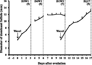
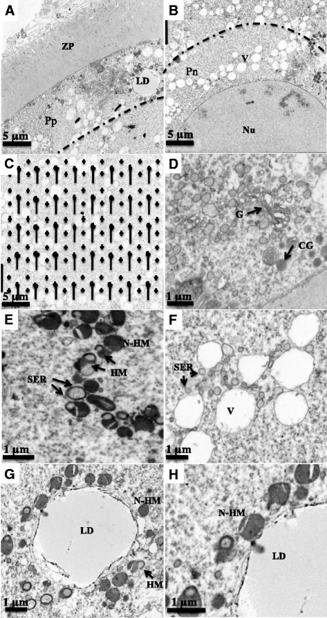
Cumulus-oocyte-complexes were collected from Hereford heifers by ultrasound-guided follicle aspiration from dominant follicles in the growing phase (n = 5; Day 0 = ovulation), static phase (n = 5), regressing phase (n = 7) of Wave 1 and from preovulatory follicles (n = 5). Oocytes were processed and transmission electron micrographs of ooplasm representing peripheral, perinuclear and central regions were evaluated using standard stereological methods.
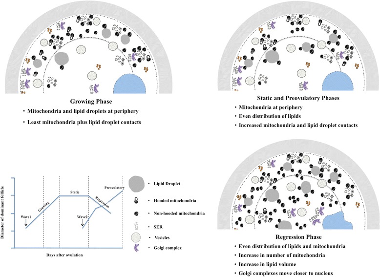
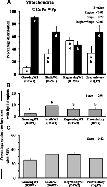
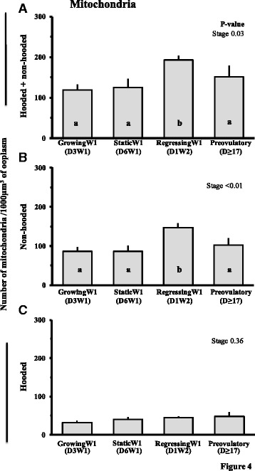
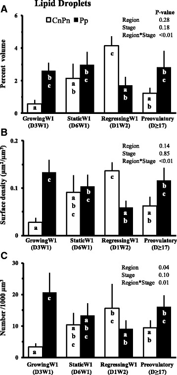
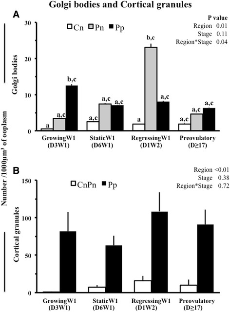
The number of mitochondria and volume occupied by lipid droplets was higher (P < 0.03) in oocytes from regressing follicles (193.0 ± 10.4/1000 μm(3) and 3.5 ± 0.7 %) than growing and preovulatory stages (118.7 ± 14.4/1000 μm(3) and 1.1 ± 0.3 %; 150.5 ± 28.7/1000 μm(3) and 1.6 ± 0.2 %, respectively). Oocytes from growing, static and preovulatory follicles had >70 % mitochondria in the peripheral regions whereas oocytes from regressing follicles had an even distribution. Oocytes from growing follicles had more lipid droplets in peripheral region than in central region (86.9 vs. 13.1 %). Percent surface area of mitochondria in contact with lipid droplets increased from growing (2.3 %) to static, regressing or preovulatory follicle stage (8.9, 6.1 and 6.2 %). The amount, size and distribution of other organelles did not differ among phases (P > 0.11).
Our hypothesis was supported in that mitochondrial number increased and translocation occurred from a peripheral to an even distribution as follicles entered the regressing phase. In addition, lipid droplets underwent spatial reorganization from a peripheral to an even distribution during the growing phase and mitochondria-lipid contact area increased with follicle maturation.
我们检验了这样一个假设,即牛卵母细胞中的细胞器在数量和空间分布上会以一种特定于卵泡发育阶段的方式发生变化。
通过超声引导下从处于生长阶段(n = 5;第0天 = 排卵)、静止阶段(n = 5)、第1波的退化阶段(n = 7)的优势卵泡以及排卵前卵泡(n = 5)中采集卵丘 - 卵母细胞复合体。对卵母细胞进行处理,并使用标准的体视学方法评估代表外周、核周和中央区域的卵质的透射电子显微镜图像。
退化卵泡中的卵母细胞中线粒体数量和脂滴所占体积高于生长阶段和排卵前阶段(分别为193.0 ± 10.4/1000 μm³和3.5 ± 0.7%,生长阶段为118.7 ± 14.4/1000 μm³和1.1 ± 0.3%;排卵前阶段为150.5 ± 28.7/1000 μm³和1.6 ± 0.2%,P < 0.03)。来自生长、静止和排卵前卵泡的卵母细胞外周区域的线粒体含量超过70%,而来自退化卵泡的卵母细胞则呈均匀分布。生长卵泡的卵母细胞外周区域的脂滴比中央区域更多(86.9%对13.1%)。与脂滴接触的线粒体表面积百分比从生长阶段(2.3%)增加到静止、退化或排卵前卵泡阶段(8.9%、6.1%和6.2%)。其他细胞器的数量、大小和分布在各阶段之间没有差异(P > 0.11)。
我们的假设得到了支持,即随着卵泡进入退化阶段,线粒体数量增加且发生了从外周到均匀分布的转位。此外,在生长阶段脂滴经历了从外周到均匀分布的空间重组,并且线粒体 - 脂滴接触面积随着卵泡成熟而增加。