Hatori Akiko, Yui Joji, Xie Lin, Kumata Katsushi, Yamasaki Tomoteru, Fujinaga Masayuki, Wakizaka Hidekatsu, Ogawa Masanao, Nengaki Nobuki, Kawamura Kazunori, Wang Feng, Zhang Ming-Rong
Molecular Imaging Center, National Institute of Radiological Sciences, 4-9-1 Anagawa, Inage-ku, Chiba 263-8555, Japan.
Department of Nuclear Medicine, Nanjing First Hospital, Affiliated to Nanjing Medical University, 68 Chanle Road, Nanjing 210006, China.
Sci Rep. 2015 Nov 27;5:17327. doi: 10.1038/srep17327.
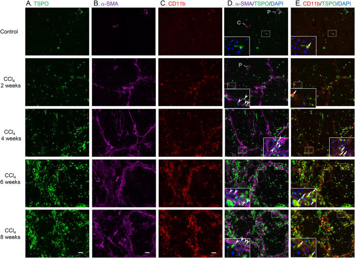
Hepatic fibrosis is the wound healing response to chronic hepatic injury caused by various factors. In this study, we aimed to evaluate the utility of translocator protein (18 kDa) (TSPO) as a molecular imaging biomarker for monitoring the progression of hepatic fibrosis to cirrhosis. Model rats were induced by carbon tetrachloride (CCl4), and liver fibrosis was assessed. Positron emission tomography (PET) with N-benzyl-N-methyl-2-[7,8-dihydro-7-(2-[(18)F]fluoroethyl)-8-oxo-2-phenyl-9H-purin-9-yl]-acetamide ([(18)F]FEDAC), a radioprobe specific for TSPO, was used for noninvasive visualisation in vivo. PET scanning, immunohistochemical staining, ex vivo autoradiography, and quantitative reverse-transcription polymerase chain reaction were performed to elucidate the relationships among radioactivity uptake, TSPO levels, and cellular sources enriching TSPO expression in damaged livers. PET showed that uptake of radioactivity in livers increased significantly after 2, 4, 6, and 8 weeks of CCl4 treatment. Immunohistochemistry demonstrated that TSPO was mainly expressed in macrophages and hepatic stellate cells (HSCs). TSPO-expressing macrophages and HSCs increased with the progression of liver fibrosis. Interestingly, the distribution of radioactivity from [(18)F]FEDAC was well correlated with TSPO expression, and TSPO mRNA levels increased with the severity of liver damage. TSPO was a useful molecular imaging biomarker and could be used to track the progression of hepatic fibrosis to cirrhosis with PET.
肝纤维化是肝脏对各种因素所致慢性肝损伤的伤口愈合反应。在本研究中,我们旨在评估转位蛋白(18 kDa)(TSPO)作为分子影像生物标志物用于监测肝纤维化向肝硬化进展的效用。用四氯化碳(CCl4)诱导建立模型大鼠,并评估肝纤维化情况。使用对TSPO具有特异性的放射性示踪剂N-苄基-N-甲基-2-[7,8-二氢-7-(2-[(18)F]氟乙基)-8-氧代-2-苯基-9H-嘌呤-9-基]-乙酰胺([(18)F]FEDAC)进行正电子发射断层扫描(PET),以在体内进行无创可视化。进行PET扫描、免疫组织化学染色、离体放射自显影以及定量逆转录聚合酶链反应,以阐明放射性摄取、TSPO水平以及受损肝脏中富集TSPO表达的细胞来源之间的关系。PET显示,CCl4处理2、4、6和8周后肝脏中的放射性摄取显著增加。免疫组织化学表明,TSPO主要在巨噬细胞和肝星状细胞(HSC)中表达。随着肝纤维化的进展,表达TSPO的巨噬细胞和HSC数量增加。有趣的是,[(18)F]FEDAC的放射性分布与TSPO表达密切相关,且TSPO mRNA水平随肝损伤严重程度增加而升高。TSPO是一种有用的分子影像生物标志物,可用于通过PET追踪肝纤维化向肝硬化的进展。