Balogh Viktoria, MacAskill Mark G, Hadoke Patrick W F, Gray Gillian A, Tavares Adriana A S
Centre for Cardiovascular Science, The Queen's Medical Research Institute, The University of Edinburgh, Edinburgh, United Kingdom.
Edinburgh Imaging, The Queen's Medical Research Institute, The University of Edinburgh, Edinburgh, United Kingdom.
Front Cardiovasc Med. 2021 Aug 17;8:719031. doi: 10.3389/fcvm.2021.719031. eCollection 2021.
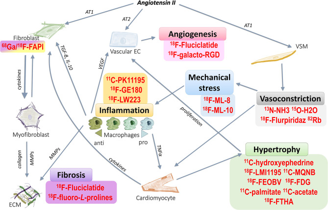
Heart failure, which is responsible for a high number of deaths worldwide, can develop due to chronic hypertension. Heart failure can involve and progress through several different pathways, including: fibrosis, inflammation, and angiogenesis. Early and specific detection of changes in the myocardium during the transition to heart failure can be made via the use of molecular imaging techniques, including positron emission tomography (PET). Traditional cardiovascular PET techniques, such as myocardial perfusion imaging and sympathetic innervation imaging, have been established at the clinical level but are often lacking in pathway and target specificity that is important for assessment of heart failure. Therefore, there is a need to identify new PET imaging markers of inflammation, fibrosis and angiogenesis that could aid diagnosis, staging and treatment of hypertensive heart failure. This review will provide an overview of key mechanisms underlying hypertensive heart failure and will present the latest developments in PET probes for detection of cardiovascular inflammation, fibrosis and angiogenesis. Currently, selective PET probes for detection of angiogenesis remain elusive but promising PET probes for specific targeting of inflammation and fibrosis are rapidly progressing into clinical use.
心力衰竭在全球范围内导致大量死亡,它可能由慢性高血压发展而来。心力衰竭可通过多种不同途径发生并进展,包括:纤维化、炎症和血管生成。在向心力衰竭转变过程中,可通过使用分子成像技术,包括正电子发射断层扫描(PET),对心肌变化进行早期和特异性检测。传统的心血管PET技术,如心肌灌注成像和交感神经支配成像,已在临床层面确立,但在评估心力衰竭时,往往缺乏对重要途径和靶点的特异性。因此,需要识别新的炎症、纤维化和血管生成的PET成像标志物,以辅助高血压性心力衰竭的诊断、分期和治疗。本综述将概述高血压性心力衰竭的关键机制,并介绍用于检测心血管炎症、纤维化和血管生成的PET探针的最新进展。目前,用于检测血管生成的选择性PET探针仍然难以捉摸,但用于特异性靶向炎症和纤维化的有前景的PET探针正迅速进入临床应用。