Hariri Omid R, Quadri Syed A, Farr Saman, Gupta Ravi, Bieber Andrew J, Dyurgerova Anya, Corsino Casey, Miulli Dan, Siddiqi Javed
Department of Neurosurgery, Arrowhead Regional Medical Center, Colton, California, United States ; Department of Neurosurgery, Institute of Clinical Orthopedics and Neurosciences, Desert Regional Medical Center, Palm Springs, California, United States.
Division of Neurosurgery, Department of Surgery, College of Osteopathic Medicine, Western University of Health Sciences, Pomona, California, United States.
J Neurol Surg Rep. 2015 Nov;76(2):e227-32. doi: 10.1055/s-0035-1560048. Epub 2015 Aug 4.
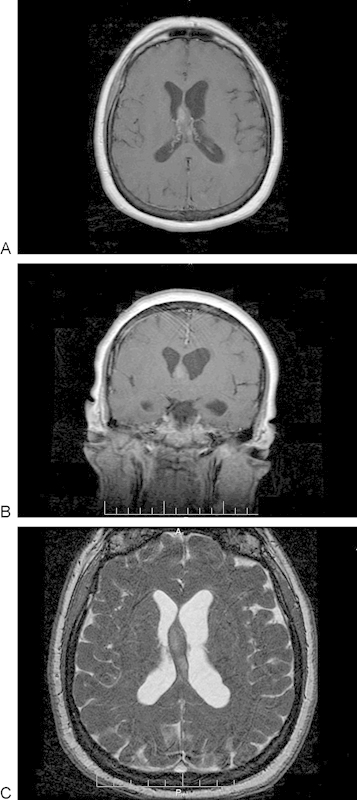
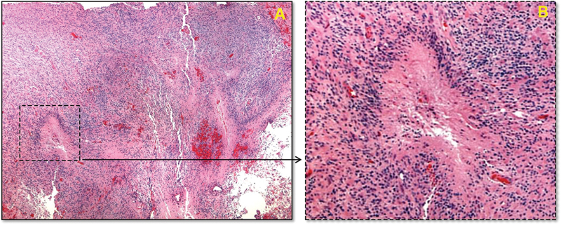
Background Glioblastoma multiforme (GBM) typically presents in the supratentorial white matter, commonly within the centrum semiovale as a ring-enhancing lesion with areas of necrosis. An atypical presentation of this lesion, both anatomically as well as radiographically, is significant and must be part of the differential for a neoplasm in this anatomical location. Case Description We present a case of a 62-year-old woman with headaches, increasing somnolence, and cognitive decline for several weeks. Magnetic resonance imaging demonstrated mild left ventricular dilatation with a well-marginated, homogeneous, and nonhemorrhagic lesion located at the ceiling of the third ventricle within the junction of the septum pellucidum and fornix, without exhibiting the typical radiographic features of hemorrhage or necrosis. Final pathology reports confirmed the diagnosis of GBM. Conclusion This case report describes an unusual location for the most common primary brain neoplasm. Moreover, this case identifies the origin of a GBM related to the paracentral ventricular structures infiltrating the body of the fornix and leaves of the septum pellucidum. To our knowledge this report is the first reported case of a GBM found in this anatomical location with an entirely atypical radiographic presentation.
多形性胶质母细胞瘤(GBM)通常出现在幕上白质,常见于半卵圆中心,表现为环形强化病灶并伴有坏死区域。该病灶在解剖学和影像学上的非典型表现具有重要意义,在该解剖位置的肿瘤鉴别诊断中必须予以考虑。病例描述:我们报告一例62岁女性,出现头痛、嗜睡加重及认知功能下降数周。磁共振成像显示轻度左心室扩张,在透明隔与穹窿交界处的第三脑室顶部有一个边界清晰、均匀且无出血的病灶,未表现出典型的出血或坏死影像学特征。最终病理报告确诊为GBM。结论:本病例报告描述了最常见的原发性脑肿瘤的一个不寻常位置。此外,该病例确定了一个与脑室旁中央结构相关的GBM起源,该结构浸润了穹窿体和透明隔叶。据我们所知,本报告是首例在该解剖位置发现的具有完全非典型影像学表现的GBM病例。