Le Sarah N, Porebski Benjamin T, McCoey Julia, Fodor James, Riley Blake, Godlewska Marlena, Góra Monika, Czarnocka Barbara, Banga J Paul, Hoke David E, Kass Itamar, Buckle Ashley M
Biomedicine Discovery Institute and Department of Biochemistry and Molecular Biology, Monash University, Clayton, Australia.
The Centre of Postgraduate Medical Education, Department of Biochemistry and Molecular Biology, Warsaw, Poland.
PLoS One. 2015 Dec 1;10(12):e0142615. doi: 10.1371/journal.pone.0142615. eCollection 2015.
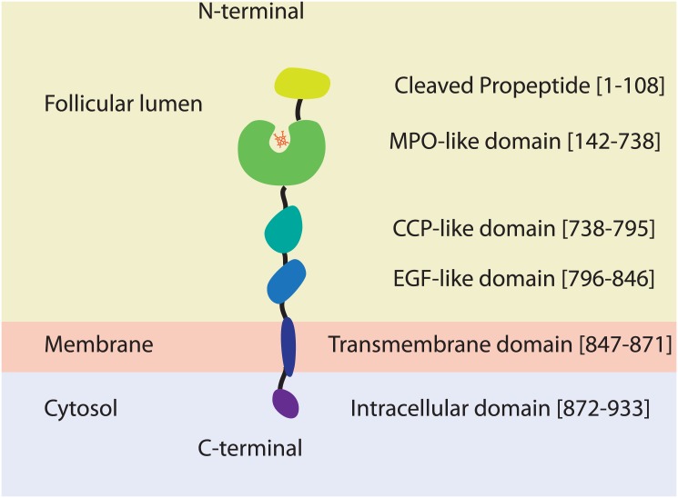
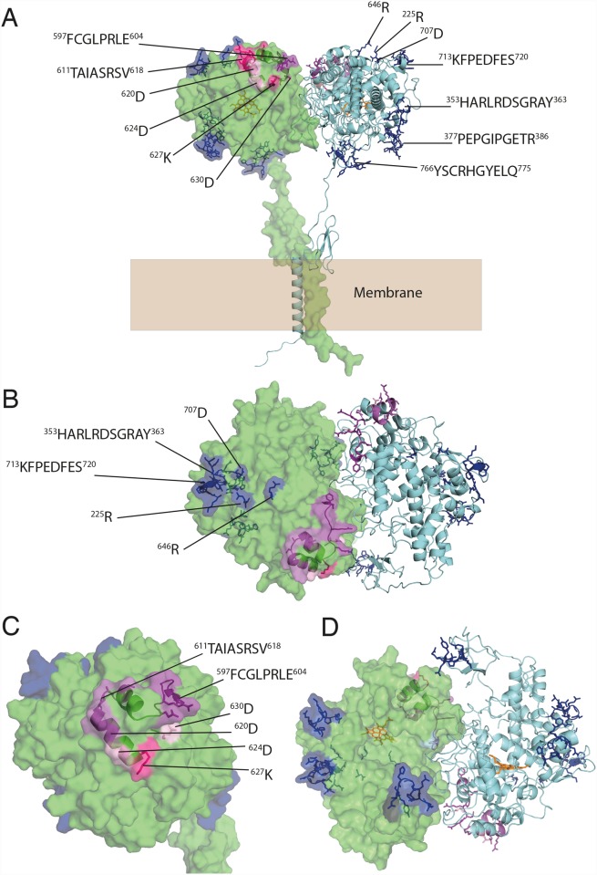
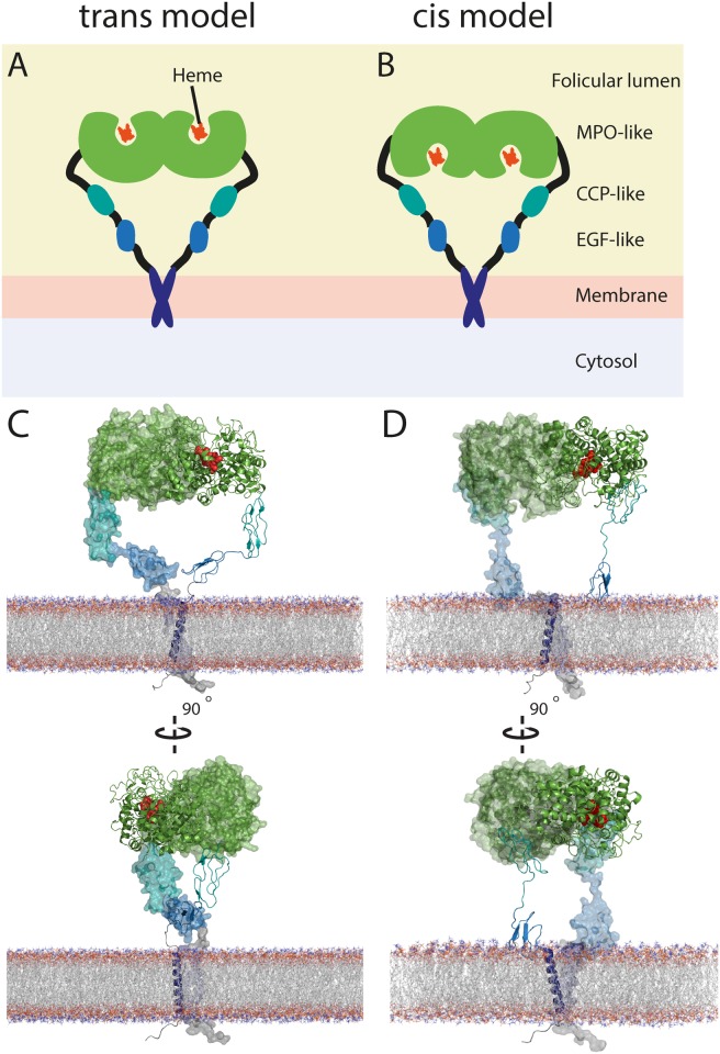
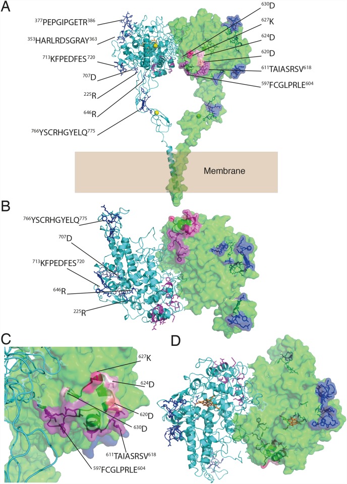
Thyroid peroxidase (TPO) catalyses the biosynthesis of thyroid hormones and is a major autoantigen in Hashimoto's disease--the most common organ-specific autoimmune disease. Epitope mapping studies have shown that the autoimmune response to TPO is directed mainly at two surface regions on the molecule: immunodominant regions A and B (IDR-A, and IDR-B). TPO has been a major target for structural studies for over 20 years; however, to date, the structure of TPO remains to be determined. We have used a molecular modelling approach to investigate plausible modes of TPO structure and dimer organisation. Sequence features of the C-terminus are consistent with a coiled-coil dimerization motif that most likely anchors the TPO dimer in the apical membrane of thyroid follicular cells. Two contrasting models of TPO were produced, differing in the orientation and exposure of their active sites relative to the membrane. Both models are equally plausible based upon the known enzymatic function of TPO. The "trans" model places IDR-B on the membrane-facing side of the myeloperoxidase (MPO)-like domain, potentially hindering access of autoantibodies, necessitating considerable conformational change, and perhaps even dissociation of the dimer into monomers. IDR-A spans MPO- and CCP-like domains and is relatively fragmented compared to IDR-B, therefore most likely requiring domain rearrangements in order to coalesce into one compact epitope. Less epitope fragmentation and higher solvent accessibility of the "cis" model favours it slightly over the "trans" model. Here, IDR-B clusters towards the surface of the MPO-like domain facing the thyroid follicular lumen preventing steric hindrance of autoantibodies. However, conformational rearrangements may still be necessary to allow full engagement with autoantibodies, with IDR-B on both models being close to the dimer interface. Taken together, the modelling highlights the need to consider the oligomeric state of TPO, its conformational properties, and its proximity to the membrane, when interpreting epitope-mapping data.
甲状腺过氧化物酶(TPO)催化甲状腺激素的生物合成,并且是桥本氏病(最常见的器官特异性自身免疫性疾病)中的主要自身抗原。表位作图研究表明,针对TPO的自身免疫反应主要针对该分子上的两个表面区域:免疫显性区域A和B(IDR - A和IDR - B)。20多年来,TPO一直是结构研究的主要目标;然而,迄今为止,TPO的结构仍有待确定。我们采用分子建模方法来研究TPO结构和二聚体组织的可能模式。C末端的序列特征与卷曲螺旋二聚化基序一致,该基序很可能将TPO二聚体锚定在甲状腺滤泡细胞的顶膜中。生成了两种截然不同的TPO模型,它们的活性位点相对于膜的方向和暴露程度不同。基于TPO已知的酶功能,这两种模型同样合理。“反式”模型将IDR - B置于髓过氧化物酶(MPO)样结构域的面向膜侧,这可能会阻碍自身抗体的接近,需要相当大的构象变化,甚至可能使二聚体解离成单体。IDR - A跨越MPO和CCP样结构域,与IDR - B相比相对分散,因此很可能需要结构域重排才能聚合成一个紧密的表位。“顺式”模型的表位碎片化较少且溶剂可及性较高,这使其比“反式”模型略占优势。在这里,IDR - B聚集在面向甲状腺滤泡腔的MPO样结构域表面,防止自身抗体的空间位阻。然而,可能仍需要构象重排以允许与自身抗体充分结合,两种模型上的IDR - B都靠近二聚体界面。综上所述,该建模强调在解释表位作图数据时需要考虑TPO的寡聚状态、其构象性质及其与膜的接近程度。